Microscopy & microtechniques
Published over 16 years ago. See the latest and most current information on Microscopy & microtechniques.
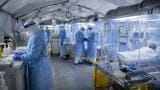
Without question, modern medicine is founded on the ability to perform pre-clinical research on diseases and their respective drug treatments. Many human disorders like cancer and infection have corresponding models in mice, providing a convenient route for pre-clinical drug and biochemical studies to take place. However, the research of these models can be cumbersome, and require large numbers of animals for study. Recent advances in small animal fluorescence imaging have dramatically increased the pace of such research, and alleviated many of the common difficulties associated with it. The tools are simple but powerful. With a given fluorescent probe or genetic reporter, a researcher is empowered to perform longitudinal imaging studies on individual cohorts of animals. This dramatically reduces the overall use of animals, while improving the statistical analysis of the data that are collected from a given experiment. The technology is safe, facile, and relatively inexpensive, and thus facilitates in vivo research at universities which may not have the imaging expertise and monetary resources of an associated medical school. Here we describe model systems in cancer that incorporate fluorescent imaging strategies, and how these methods are improving and enhancing research in this critical field.
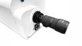
Fluorescence imaging of living specimens may be undertaken at a range of magnification. The core concept at each level is to use filtered light to excite fluorochromes within the animal, with the emitted light captured by a
charge coupled device (CCD) with appropriate emission filter. An animal may be imaged at the macroscopic level to detect fluorescent signals emanating from all tissues. This is typically referred to as “whole animal fluorescence imaging” and has the advantage of providing a rapid snapshot of events taking place within the context of the whole body. Next, fluorescence imaging may also be performed at the single cell level within animals. This is referred to as ‘intravital microscopy (IVM)’ and is a powerful tool for directly visualising individual cells and their processes in living animals. IVM imparts a unique advantage to fluorescence based methods over other optical modalities like bioluminescence, which do not have the spatial resolution to image at the cell and sub-cellular level. The use of optical wavelength light makes the technology safe and easy to use, thus enabling a multitude of applications for cancer imaging.
ILM Guide 2026/27


.jpg)