Microscopy & Microtechniques
Report on the Exploration of Living Cells using Nanoscale and Single Molecule Techniques through the Application of Scanning Probe Microscopy
Jun 24 2015
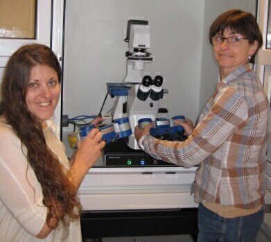
Dr Yves Dufrêne is a senior research fellow who is leading the Nanobio Team at UCL, Université catholique de Louvain. His research is at the cross-roads of nanotechnology and microbiology where the goal is to gain insight into the nanometer-scale surface architecture and molecular interactions of microbes. Two main topics are: understanding the spatial organisation of the cell wall and deciphering the molecular mechanisms of cell adhesion.
Looking more deeply at his work, Dr Dufrêne said: “the completion of our multidisciplinary research requires a novel, state-of-the-art AFM for the nanoscale analysis of live cells hence our choice of the NanoWizard® from JPK Instruments. This is equipped with a series of advanced capabilities that are essential for the project. First, the combination of AFM with a high-quality inverted optical microscope will allows us to optically guide AFM analyses (selection of a cell of interest, followed by targeted AFM measurements), therefore improving dramatically the throughput and quality of single-cell analyses. In addition, this AFM-optical microscopy platform will make it possible to directly correlate AFM topographic images with fluorescence images and to prepare living cell probes for single-cell force spectroscopy (SCFS) measurements. Secondly, the new high-speed module will complement traditional topographic imaging of bacterial cell surfaces by providing a more dynamic view of the cell surface (cell surface organisation, relaxation and turnover; cell wall remodelling in response to drugs). We believe this project is a unique opportunity to push the limits of high-speed AFM in microbiology. Third, an advanced single-molecule force spectroscopy (SMFS) package will enable us to image and manipulate, at high resolution, single cell surface constituents. This package will include the force-clamp mode, which will provide novel insights into the adhesive and mechanical properties of the adhesins. Fourth, the newly developed multiparametric imaging mode (also named quantitative imaging, QI™) will allow us to dramatically increase the spatiotemporal resolution of SMFS-based imaging, and to correlate the localisation of single cell surface constituents with local changes in adhesion and elasticity. Fifth, the unique cell adhesion force module (CellHesion™ module) will guarantee reliable, state-of-the art single-cell force spectroscopy (SCFS) measurements, and will permit, for the first time, long-range z-spectroscopy (up to 100 µm), thereby enabling to unravel molecular interactions that were not accessible before.”
Digital Edition
Lab Asia 31.2 April 2024
April 2024
In This Edition Chromatography Articles - Approaches to troubleshooting an SPE method for the analysis of oligonucleotides (pt i) - High-precision liquid flow processes demand full fluidic c...
View all digital editions


Events
Apr 28 2024 Montreal, Quebec, Canada
May 05 2024 Seville, Spain
InformEx Zone at CPhl North America
May 07 2024 Pennsylvania, PA, USA
May 14 2024 Oklahoma City, OK, USA
May 15 2024 Birmingham, UK


