-
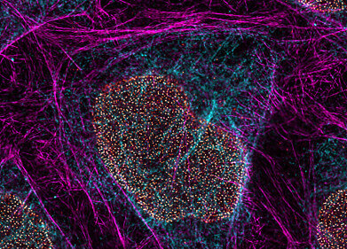
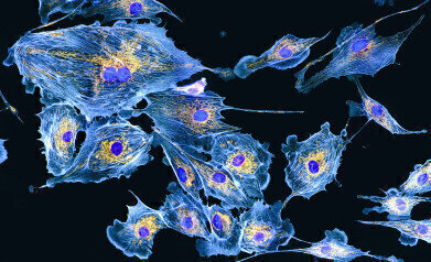 Endothelial cells, from the lining of blood vessels in the lungs, are shown in exquisite detail by the technique of immunofluorescence microscopy. The sample has been treated with antibodies that bind to specific proteins within parts of the cell and with dyes that bind to these specific antibodies. Each dye has its own fluorescence colour, so when illuminated by ultraviolet light the colours show the location of each target protein. Here, purple dye has attached to proteins in the cell nuclei, blue dye shows actin filaments that form the structural cytoskeleton and yellow shows the mitochondria that generate energy for the cell. Credit Dr Torsten Wittman
 -
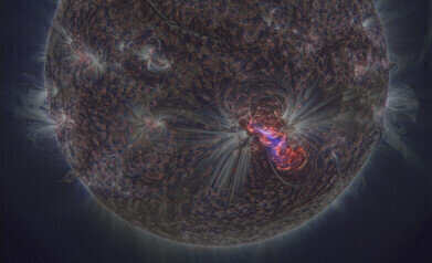 This dramatic image, captured by the orbiting Solar Dynamics Observatory, shows one of the most active sunspot groups observed in the last few decades. Solar active region AR2192 was the source of six solar flares of the largest class (X-class) in October 2014. This image was gathered in extreme ultraviolet wavelengths - 193 ångstroms in blue, 171 ångstroms in white and 304 ångstroms in red (one ångstrom is a ten billionth of a metre). The UV light comes from highly ionised helium and iron atoms in the Sun’s outer atmosphere, and shows very clearly how the atoms congregate along the intense magnetic field lines. The rest of the Sun’s surface, so bright to our eyes, appears dark at such extreme wavelengths. This exceptionally sharp image was created by processing the raw data using special software called NAFE. Credi: NASA/SDO/AIA, processing by Miloslav Druckmuller
 -
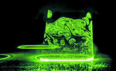 The very things that make glass for smartphones useful - their hardness and thinness - make them almost impossible to cut to shape by conventional means. Extremely fast lasers make the job possible. By scanning across cut lines several times using superfast pulses of light, the laser can cut the strongest types of glass in any shape and with a smooth edge contour. This removes the need for lengthy shaping and polishing operations. The laser produces pulses of a few tens of picoseconds (million millionths of a second) duration at a rate of tens of millions of pulses per second. This prevents localised heat build-up which could cause stress fractures in the glass .Credit:Fraunhofer Institute for Laser Technology ILT, Aachen, Germany / Volker Lannert
 -
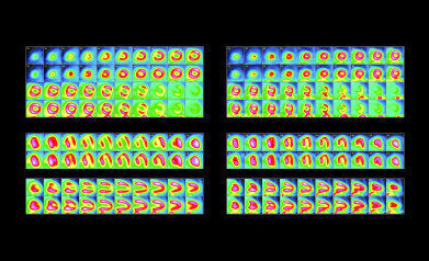 Myocardial Diffusion Imaging shows a patient’s heart before and after surgery. To obtain the images, the patient was injected with a chemical that binds to active myocardial (heart muscle) tissue, and includes rubidium-82, a radioactive marker. This emits gamma rays, so a gamma camera can make multiple scans to create a 3D map of the active heart muscle. Here the upper block shows slices from top to bottom of the heart, the middle block shows slices from back to front and the lowest block slices from the centre of the body outward. Alternate rows show the heart stressed and resting. In the array of images at left there are areas of significant missing activity, particularly in the stress images. This is due to a 99% blockage of the left anterior descending coronary artery. The blockage was treated by implanting a stent (mesh-like tube) at the blockage site.Credit:Dr Eric Hong Cho Tek, EH Heart Specialist Private Limited, Singapore
 -
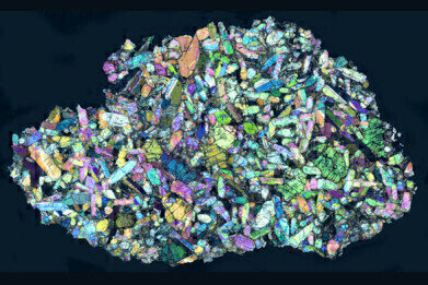 A view of a thin section through a Martian meteorite found at Nakhla, Egypt, in 1911. The sample is lit from below with polarised light, then another polarising filter is placed between the sample and the eyepiece. Different minerals rotate polarised light by different amounts creating characteristic colours, so the technique allows scientists to determine the minerals present. This rock is a clinopyroxinite consisting largely of augite and olivine with smaller amounts of other minerals. It is thought that this rock formed around 1.38 billion years ago on Mars. It was blasted from the surface of the planet by a huge impact event, meandering its way through the Solar System for an unknown time before hitting the Earth’s atmosphere and breaking up. This sample is approximately 13mm across. Credit:© Natural History Museum London BM1913, 2642 and the Open University Virtual microscope
Microscopy & Microtechniques
Incredible Images Launch International Year of Light 2015
Jan 29 2015
Images from around the world defining what we see at the largest and smallest scales, including some that are normally invisible to the human eye, are just part of a new photography exhibition which opened in London and is currently on a national tour.
Organised by the Royal Photographic Society (RPS) with support from the Royal Astronomical Society (RAS) and the Science and Technology Facilities Council (STFC), Curated by RPS Fellow Gary Evans, Light Works celebrates the International Year of Light 2014 and on a virtual journey through the electromagnetic spectrum from gamma rays to radio waves, it explore ways of looking into our own bodies and the world around us. The stunning images will offer something to everyone, regardless of their age or level of knowledge.
Gary Evans said: “With images so ubiquitous in the world today it was clear that the Royal Photographic Society needed to do something special to celebrate the International Year of Light. A ‘walk through the electromagnetic spectrum’ was the way to go, the hard part was settling on 50 images that not only have a rich visual impact, but also a story to tell.”
Dr Andrew Taylor, Director of STFC’s National Laboratories, said, “At STFC we use light every day in many different ways. For instance, we use intense beams of light to develop new methods of diagnosing medical conditions, to investigate new materials for energy production and storage, and to see into the depths of space. We are very pleased to be able to help bring this exhibition of photographs to people in the UK for the start of this International Year of Light.”
The many contributors of images have been generous in their support and collaboration to make the exhibition possible.
Dr Michael Pritchard, Director General of the RPS, commented: “Photography has played an important role in documenting the visible and invisible world since the 1840s. Modern imaging techniques using visible and invisible light – as Light Works shows - can provide us with a view into the infinitesimal small and the gigantic. More than this they provide us with beautiful images that inspire and excite us”
Professor Martin Barstow, President of the Royal Astronomical Society, said, “It is wonderful to see such incredible images from astronomy being presented in this way to the public. However, what was enlightening about this exhibition was experiencing the beauty and power of the images taken at the opposite scale to astronomical images or taken within different parts of the electromagnetic spectrum. These images offer everyone a way to see the world around us in a completely fresh light.”
To find out where the exhibition is or view the images visit: www.rps.org/exhibitions-and-competitions/current-exhibitions/rps-light-works-exhibition

Digital Edition
Lab Asia 31.2 April 2024
April 2024
In This Edition Chromatography Articles - Approaches to troubleshooting an SPE method for the analysis of oligonucleotides (pt i) - High-precision liquid flow processes demand full fluidic c...
View all digital editions


Events
Apr 28 2024 Montreal, Quebec, Canada
May 05 2024 Seville, Spain
InformEx Zone at CPhl North America
May 07 2024 Pennsylvania, PA, USA
May 14 2024 Oklahoma City, OK, USA
May 15 2024 Birmingham, UK


