Microscopy & Microtechniques
RISE Microscopy for Correlative Raman-SEM Imaging Launched at Analytica 2014
Mar 26 2014
Tescan Orsay Holdings and WITec GmbH jointly launched RISE Microscopy at Analytica 2014.
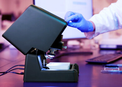
RISE Microscopy is a novel correlative microscopy technique which combines confocal Raman Imaging and Scanning Electron (RISE) Microscopy within one integrated microscope system. This unique combination provides clear advantages for the microscope user with regard to comprehensive sample characterisation. The RISE Microscope enables for the first time the acquisition of SEM and Raman images from the same sample area and the correlation of ultra-structural and chemical information with one microscope system.
Both analytical methods are fully integrated into the RISE Microscope. Between the different measurements an extremely precise scan stage automatically transfers the sample inside the microscope’s vacuum chamber and re-positions it. The integrated RISE software carries out the required parameter adjustments and instrument alignments. The acquired results can then be correlated and the Raman and SEM images overlaid. “RISE Microscopy enables unprecedented opportunities for the most comprehensive ultra-structural and molecular sample analyses,” explained Dr Olaf Hollricher, CEO and Director R&D at WITec “The novel RISE Microscope is another striking example of WITec’s enormous innovative strength. It fulfils all requirements of an outstanding, correlative microscopy technique and will convince the Raman as well as the SEM community.”
TESCAN and WITec arranged worldwide sales and after-sales cooperation for the RISE Microscope to take advantage of the synergy effects of both companies.
The RISE Microscope provides all functions and features of a stand-alone SEM and a confocal Raman microscope. Both SEM and Raman are high-resolution imaging techniques with sub-nanometer and diffraction limited 200 - 300 nanometer resolution, respectively. In Raman imaging mode the sample can be scanned through a range of 250 µm x 250 µm x 250 µm. RISE Microscopy pairs ease-of-use with exceptional analysing benefits and is therefore suited to a large variety of applications such as nanotechnology, materials science, and life science.
Digital Edition
Lab Asia 31.2 April 2024
April 2024
In This Edition Chromatography Articles - Approaches to troubleshooting an SPE method for the analysis of oligonucleotides (pt i) - High-precision liquid flow processes demand full fluidic c...
View all digital editions


.jpg)
Events
Apr 22 2024 Marrakech, Morroco
Making Pharmaceuticals Exhibition & Conference
Apr 23 2024 Coventry, UK
Apr 23 2024 Kintex, South Korea
Apr 23 2024 Seoul, South Korea
Apr 24 2024 Jakarta, Indonesia


