-
 Professor Steve Williams, Centre for Imaging Science, Manchester University
News & Views
MRI Scanner Assists Manchester’s Stem Cell Safety Hub
Feb 25 2015
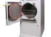
Researchers at the University of Manchester’s Centre for Imaging Science are using a newly installed MRI pre-clinical imaging system from MR Solutions in their work to develop methods and technology for assessment of regenerative medicine therapies. The team, led by Professor Steve Williams aims to provide a framework for the most appropriate label and imaging technology to use for monitoring the biodistribution and behaviour of transplanted cells in well-characterised disease models using innovative nanochemistry and molecular imaging. Using such robust assessment methods will assist those developing novel therapies by accelerating the transfer of successful drug candidates into a clinical setting.
The new MRS 3000 system, funded by the UK Regenerative Medicine Platform (http://www.ukrmp.org.uk/hubs/) was chosen for its soft tissue contrast and molecular imaging capability and with its cryogen-free magnet configuration and well-contained stray magnetic field can also be used safely in most laboratory situations.
“We wanted a system that we could locate in the same laboratory as an existing PET scanner; as this MRI instrument requires no cryogens, has a small footprint and almost no stray field we can carry out multi-modal imaging in an ordinary laboratory environment,” Professor Williams told Labmate UK & Ireland.
The research being conducted at Manchester is part of the ‘Stem Cell Safety Hub’ a multi-institutional collaboration led by Liverpool University with Manchester, UCL and Edinburgh Universities, with core teams composed of chemists, pharmacologists, imaging experts, stem cell biologists and clinicians.
“The Safety Hub at Manchester is studying the safety and efficacy of stem cells in established disease models by methods that both track where the stem cells are delivered to and also the response of the host tissue to their presence. Working with chemists, clinicians, cell biologists and statisticians from Manchester and Liverpool we are using imaging to characterize structural and functional consequences of kidney damage so we can subsequently determine if stem cell therapy will mitigate this damage. In combination with other non-invasive imaging techniques we can also help to identify the mechanisms of damage and repair, ” Professor Williams added.


Digital Edition
Lab Asia 31.2 April 2024
April 2024
In This Edition Chromatography Articles - Approaches to troubleshooting an SPE method for the analysis of oligonucleotides (pt i) - High-precision liquid flow processes demand full fluidic c...
View all digital editions


Events
Apr 28 2024 Montreal, Quebec, Canada
May 05 2024 Seville, Spain
InformEx Zone at CPhl North America
May 07 2024 Pennsylvania, PA, USA
May 14 2024 Oklahoma City, OK, USA
May 15 2024 Birmingham, UK


