-
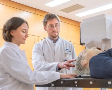 Mária Martišíková (left), the project leader from Heidelberg University Hospital and German Cancer Research Center (DKFZ), and DKFZ researcher Laurent Kelleter. (Image Credit: Heidelberg University Hospital / H.Schroeder)
 -
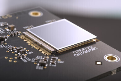 The Timepix3 chip developed at CERN (Credit: CERN)
Research News
Small CERN detector could help radiotherapy precision of head tumours
Mar 15 2024
Particle detectors similar to those used by physicists at CERN have now also found a role in a new imaging device now being tested on patients by scientists from the German National Center for Tumor Diseases (NCT), the German Cancer Research Center (DKFZ), and the Heidelberg Ion Beam Therapy Center (HIT) at Heidelberg University Hospital. Supplied by Czech company ADVACAM the device, which includes a small Timepix3 pixel detector developed at CERN, enables head and neck tumours to be closely monitored during ion radiotherapy, making them easier to target while helping to limit the treatment’s side effects.
"One of the most advanced methods for treating head and neck tumours involves irradiation with ion beams. This has one unique feature: it can be precisely tailored to the depth inside the human head where the particles should have the maximal effect”, explained Mária Martišíková, the head of the DKFZ team.
Ion radiation, like other types of irradiation, also has the drawback of affecting healthy tissue areas around the tumour and precise targeting of tumours is particularly challenging in the brain, where damage to the optic nerve or a patient’s memory are possible. X-ray computed tomography (CT) scan images taken before treatment essentially provide a map to target the tumour with ion beams; however further complications can arise during therapy as changes in the skull may evolve that alter the target and until now, physicians have lacked a reliable tool to alert them in case of such a change in the brain.
The new device now offers the potential for improving the navigation of the ion beams inside the head by tracking the secondary particles that are created when ions pass through it.
"Our cameras can register every charged particle of secondary radiation emitted from the patient's body. It's like watching balls scattered by a billiard shot. If the balls bounce as expected according to the CT image, we can be sure we are targeting correctly. Otherwise, it's clear that the 'map' no longer applies. Then it is necessary to replan the treatment," said Lukáš Marek from ADVACAM.
"We hope the new device will show us how often and where the tumour changes occur. It will allow us to reduce the overall irradiated volume of tissue, saving healthy tissue and reducing the side effects of radiotherapy. We will also be able to apply higher doses of radiation to the tumour" adds Martišíková.
“When we started developing pixel detectors for the LHC we had one target in mind – to detect and image each particle interaction and thereby help physicists to unravel the secrets of Nature at high energies. The Timepix detectors were developed by the multidisciplinary Medipix Collaborations whose aims are to take the same technology to new fields. Many of those fields were completely unforeseen at the beginning and this application is a brilliant example of that,” added Michael Campbell, Spokesperson of the Medipix Collaborations.
More information online


Digital Edition
Lab Asia 31.2 April 2024
April 2024
In This Edition Chromatography Articles - Approaches to troubleshooting an SPE method for the analysis of oligonucleotides (pt i) - High-precision liquid flow processes demand full fluidic c...
View all digital editions


.jpg)


Events
May 05 2024 Seville, Spain
InformEx Zone at CPhl North America
May 07 2024 Pennsylvania, PA, USA
May 14 2024 Oklahoma City, OK, USA
May 15 2024 Birmingham, UK
May 21 2024 Lagos, Nigeria


