Microscopy & Microtechniques
What is Multi-Pass Microscopy?
Oct 04 2016
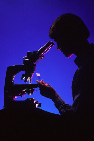
In microscopy, there is always a call for more sensitive and more accurate microscopes. Developments in this area allow scientists to look at a level beneath what they thought was possible, opening up a whole new world of potential research. And physicists at Stanford University have done just that. They’ve developed a new, more sensitive method of microscopy. So how does it work?
Lights, camera, action
One of the problems for scientists using microscopes is a dilemma with lighting. They’re trying to examine delicate biological samples. So to avoid damaging them, they have to work in a low-lighting environment. This is where the problem lies, because much like a picture you might take yourself, the low lighting causes the images to suffer.
For a normal photo, this means you can’t see the details very well – and it’s not much different for microscopy. The more intricate details like internal structure and proteins of the specimens are hard to examine in these images. This effect, where low lighting causes a grainy quality and lack of detail, is known as ‘shot noise’.
Reducing shot noise
According to the Stanford scientists, there is a way to get around shot noise, namely multi-pass microscopy. It’s a way of reusing the tiny light particles called photons. In microscopy, the photons hit the sensor, which enables a picture of the sample to be captured. However, there is a way to recycle these photons. By recapturing the same image and passing the same light through multiple versions of this image, the scientists have been able to capture more detail.
“You first take an image of the specimen, you then illuminate it with an image of itself, and the image you get, you again send back to illuminate the sample. This leads to contrast enhancement,” explains Brannon Klopfer, a Stanford graduate involved in the development. “While multi-passing builds up the signal in your image, the noise is hardly affected.”
Multi-pass electron microscopy
At the moment this idea has only been applied to optical microscopy. However, the team are working on a method of multi-pass electron microscopy. This could improve the detailed imaging of DNA and single proteins. Another innovation in electron microscopy is the addition of temporal information.
It’s something that is largely missing in regular micrographs, but there is a solution. Taking a series of snapshots creates a flip-book effect, which can help to characterise membrane trafficking events that happen over short periods of time. This development, and how it can improve research, is explored in ‘Flash-and-freeze Electron Microscopy – Adding Motion to Electron Micrographs’.
Digital Edition
Lab Asia 31.2 April 2024
April 2024
In This Edition Chromatography Articles - Approaches to troubleshooting an SPE method for the analysis of oligonucleotides (pt i) - High-precision liquid flow processes demand full fluidic c...
View all digital editions


Events
Apr 28 2024 Montreal, Quebec, Canada
May 05 2024 Seville, Spain
InformEx Zone at CPhl North America
May 07 2024 Pennsylvania, PA, USA
May 14 2024 Oklahoma City, OK, USA
May 15 2024 Birmingham, UK


