Mass spectrometry & spectroscopy
Published over 3 years ago. See the latest and most current information on Mass spectrometry & spectroscopy.
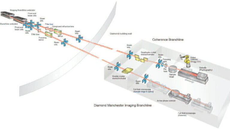
The high penetration capability of X-rays enables studies of matter close to its original state, revealing the relation between structure and functionality, accounting chemical states and compositions and X-ray spectroscopy is a powerful tool for the determination of local atomic structure in materials not characterised by crystalline order. This article looks at how the highly brilliant X-rays available at Diamond Light Source, the UK’s national synchrotron radiation source, provide extended capabilities for imaging structures across different length scales and exploring a multitude of recording modes. Processes can be recorded due to the high intensity of the radiation and chemical information can be extracted by the signature of the X-ray absorbing constituents of the component. It will explore the capabilities of imaging and tomography with high spatial, temporal and chemical resolution using this unique energy source. All the results have been achieved at the Diamond beamline I13L, which consists of two independent branches. One branch is focussed on real-space radiography and tomography, the second branch explores techniques operating in reciprocal space. The operational X-ray energy is typical 6-30keV and the resolution in the micro- to nano-range.
Figure 1. Overview of the I13L operational branches.
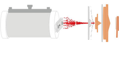
Synchrotron radiation carries a number of unique properties, one of them is a high degree of coherence. It enables exploration of the wave property of light as the observation of the wave deformation and propagation is a sensitive tool for studying soft matter. In the near-field, the distance between the sample and detector is relatively close and the edges of weakly absorbing structures are significantly enhanced. This is especially the case for bio-medical research tissue such as muscles, cells, cartilage or tendons which are rendered visible and together with more absorbing structures such as bone provide a complete picture of complex structures. For example, a sample of a mouse knee under realistic conditions can be studied, see Figure 2.
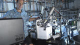
Access to synchrotron facilities is limited with a competitive peer-reviewed application process for beamtime. During the COVID period we implemented a new system allowing easy access and remote operation which significantly increased the number of specimens that could be measured. For changing samples an industrial robot arm has been installed which has currently increased sample throughput to 300 samples per day and this will be further increased with the upgrades planned for Diamond.
The decoding of the evolution of insects, their phenotype and function is a huge challenge being addressed by the National History Museum team in collaboration with Diamond. Besides enhanced recording capabilities, a workflow for data handling and analysis is required.
Figure 3. Examples of surface rendered Mycetophagidae (hairy fungus beetle: sample courtesy A. Goswami, National History Museum London)
At the I-13 beamline we cover three orders of magnitude in length-scales from micro- meters to the nano-meter range. This capability is necessary for a holistic approach to investigation: structures repeat themselves across different length scales or the cause for a macroscopic observed phenomenon relates to a microscopic cause. Typical applications require a ‘zoom-in’ capability when for example, studying the structure of the brain to the level of neurons and connectomes, crack propagation in materials, and degradation cycles of modern battery anodes to name just a few cases.
With the appearance of optics suited for the X-ray regime, it has become possible to implement schemes and instruments similar to those known from visible light optics. We have built a full-field X-ray microscope achieving sub-100nm resolution using Fresnel Zone Plates (FZP) and a special condenser optic, called beam shaper. Zernike-type microscopy is also now possible for weakly absorbing samples.
Resolution beyond the limitations of detectors and X-ray optics is achieved with imaging in reciprocal space. The diffraction pattern in the far field and the sample under coherent illumination are reciprocal (relate via the Fourier transform), in other words the large pattern on the detector corresponds to small features in the sample. A new technique, named Ptychography, helps to reconstruct phase and amplitude reliably, scanning with a focussed beam across the sample. Spectro-microscopic data is recorded as by-product, adding a fluorescence detector to the setup.
Highest spatial resolution can be used for deciphering many things. For example, the astonishing properties of moth wings. Some species have the outstanding capability of either absorbing or even transmitting sound waves, protecting them from predators such as bats. In our study we correlate the mechanical structure with sound wave simulations, learning about some of the wonders of nature.
The combination of detection modes enables relating physical and chemical structure with the functionality of materials. As an example, we illustrated the multi-modal study on particles from Fukushima fallout. By combining the different data channels a forensic study of the accident is possible. The formation of glassy material is related to a rapid cooling event, such as a blow of a container or building, and the chemical elements associated with this phase permits deducting the possible location of the blast and the series of events happening after the accident.
Figure 4. Fukushima particle - Multi-modal study on particles from Fukushima fallout (scale bar=100µm). Combining different data channels helps on a forensic study of the accident. [6].
Spectroscopic imaging is an essential tool for learning about elements and their chemical states, At Diamond a number of beamlines dedicated to spectro-microscopic imaging such as I14, I18 or I08, can study a significant part of the periodic table elements.
The branches of the I13 beamlines operate principally in two modes: either measuring the sample’s absorption or fluorescence. For the absorption case the incident X-ray photon energy is scanned around the X-ray absorption edge of a specific element, either to identify it (dual-energy) or to study its chemical state in more detail (XANES). With coherent imaging techniques such as ptychography or grating interferometry, it is now possible to determine both, phase and amplitude. Similar concepts as in visible light spectroscopy (IR/Raman) apply to relate phase and amplitude, maximising the information output.
Modern X-ray detectors have the capability to count single X-rays and to distinguish their energy to a level of >~100eV. This provides the opportunity of element sensitive detection using broadband radiation without repeating scans at different X-ray photon energies. Figure 5 illustrates a test experiment with an energy selective detector, identifying elements in a single detector scan.
Figure 5. Element-sensitive ptychography. Test sample consisting of Ni and Au grid (left), the width of the bar is 30µm. Data is recorded with a single scan using an energy selective detector and subtracting the channels around the relevant edges.
Synchrotrons are moving forward, preparing machine upgrades that will allow new possibilities for science. For example, Diamond’s planned upgrade to Diamond-II from 2027 involving a new electron storage ring, will not only increase brightness and coherence by a factor of 70, but also enhance beam quality and stability through new X-ray optics and instrumentation, state-of-the-art sample delivery, and manipulation through the development of optimised sample environments. Progress in accelerator technology means Diamond-II will offer the scientific community in academia and industry the opportunity to exploit these much brighter beams and an increased coherence over a large energy range on all Diamond’s beamlines. The coherent fraction of the beam to be used will increase by about hundred times, the flux on the imaging branch will be tenfold augmented with a new light-generating device. Multi-scale imaging will receive a significant boost with this transformational upgrade which will enable a huge expansion of UK science capabilities and provide access to new science.
The I13 team (D. Batey, A. Bodey, S. Marathe, S. Kachkanov, P. Li, K. Jakata, L. Turpin) is acknowledged, including the support teams (K. Wanelik, H. Shorthouse).
Thanks to the team of M. Holderied for collaborating on imaging moths.
R. Ziesche (HZB, Berlin), P. Shearing and M. Johnson (UCL, London) are collaborating on batteries and are acknowledged for sharing preliminary results.
1. C. Rau, Synchrotron Radiation News 30. 19 – 25, (2017), 10.1080/08940886.2017.1364530
2. https://www.diamond.ac.uk/Instruments/Imaging-and-Microscopy/I13.html
3. K. A. Staines, K. Madi, S. M. Mirczuk, et al., Arthritis & Rheumatology, 68 (4), 880–891, (2016), 10.1002/art.39508
4. K. Madi, K. A. Staines, B. K. Bay, et al., Nature Biomedical Engineering 2. (2019), 10.1038/s41551-019-0477-1
5. C. Rau, S. Marathe, A. J. Bodey, et al., SPIE Optical Engineering + Applications, 2021, 10.1117/12.2598470
6. P. G. Martin, M. Louvel, S. Cipiccia, et al., Nature Communications (2019), 10. 2801, 10.1038/s41467-019-10937-z
7. F. Brun, V.. Di Trapani, D. Batey, et al., Scientific Reports 10 (2020), 10.1038/s41598-020-63161-x
8. D. Batey, S. Cipiccia, F. Van Assche, et al., Scientific Reports 9. (2019), 10.1038/s41598-019-48642-y
ILM 51.5 July 2026


.jpg)


-(1).jpg)

.jpg)

.jpg)