Mass spectrometry & spectroscopy
Published over 13 years ago. See the latest and most current information on Mass spectrometry & spectroscopy.
Phosphorylation is a reversible post-translational modification (PTM) of proteins. A phosphate group is added to a serine, threonine or tyrosine residue via a covalent bond. Phosphorylation of proteins is important in biological molecules as it plays a role in various pathways including cell signalling and metabolism, amongst other activatory or inhibitory roles. However, atypical phosphorylation can be detrimental and has been linked to cancer, Alzheimer’s disease and a variety of other illnesses. [1, 2] For these reasons it is important that the detection and localisation of phosphorylated residues is accessible.
Tandem mass spectrometry (MS/MS) allows an in depth chemical analysis of various molecules by fragmenting ions. Collision induced dissociation (CID) is a slow heating technique that fragments the precursor ion using a low energy pathway. The low energy causes the weak peptide (CO-N) bond to break, forming b and y ions. CID of phosphorylated peptides often results in the loss of either 98 Da (phosphoric acid) or combined loss of 80Da and 18Da (phosphate and water). CID therefore allows us to identify the presence of a phosphorylation within the peptide; however it often fails to locate the exact residue that is phosphorylated.
Syka et al. [3] introduced Electron Transfer Dissociation (ETD) in 2004 as an additional fragmentation technique. ETD fragmentation is similar to ECD. [4] ETD fragments multiply charged cations by transferring electrons to them and forming charge-reduced radical ions. [4] The N-Cα bond is broken forming c and z• ions, and PTMs are preserved on the backbone. [5]
This is advantageous over CID as it increases the possibility of accurately localising the PTM sites. This also means that isobaric phosphopeptides can be identified. ETD fragmentation is random and non-selective, therefore allowing for a higher peptide sequence coverage than selective processes such as CID. [6]
A problem associated with ETD is the failure to completely fragment doubly-charged ions. [6] Unlike CID, ETD does not break the non-covalent bonds, this causes improper separation of the fragments, producing large charge reduced species and spectra which contain few fragment ions. Coon et al. overcame this problem with the introduction of supplemental activation ETD; the charge reduced ion is collisionally activated which allows separation of the fragments and more informative spectra. [7] Coon et al observed that using supplemental activation increased the sequence coverage and enhanced peptide fragmentation when compared to standard ETD. [8, 9]
High-field asymmetric waveform ion mobility spectrometry (FAIMS) separates ions in the gas phase at atmospheric pressure on the basis of their mobility at changing electric fields. [10] A voltage is applied via an asymmetric waveform causing the ions to oscillate between two electrodes. Each ion will have its own mobility towards the electrodes, however to exit from the FAIMS device the ion must be ‘balanced’ between them.
To ‘balance’ the ions, a compensation voltage (CV) is applied. Each ion has a range of CV where it is transmitted into the mass spectrometer. The CV therefore is used to define which ions are transmitted, thereby separating them. [11] FAIMS is not based solely on mass to charge ratio so it can therefore be used to separate isobaric species which is useful in phosphopeptide analysis.
Reid et al. have previously identified the gas phase relocation of a phosphate group on a peptide within CID. [12] Phosphate relocation can become a real problem to protein analysts as it leads to inaccurate localisation of the phosphorylation site.
The aim of this work is to identify the possibility of gas-phase phosphate relocation within saETD and FAIMS-ETD analysis and determine the optimum conditions in LTQ Orbitrap Velos saETD and FAIMS where this is not observed. We have varied the supplemental activation energy and the dispersion voltage applied to FAIMS and observed the differences in fragmentation of a set of 9 synthetic peptides.
Method
The following peptides were synthesised by Alta Biosciences (Birmingham, UK):
1. APLsFRGSLPKSYVK
2. APLSFRGsLPKSYVK
3. APLSFRGSLPKsYVK
4. NTNEyTEGPTVVPR
5. NTNEYtEGPTVVPR
8. APLSFLGSLPKsYVK
9. APLsFLGSLPKSYVK
11. LFtGHPESLER
12. LFTGHPEsLER
s is phosphoserine, t is phosphothreonine, y is phosphotyrosine
The peptides were re-suspended in water and made up to a 2µM concentration with a solution of 70% methanol, 30% water and 2% formic acid. Peptides were directly infused by use of an Advion Nanomate Triversa and then subject to ETD (100 ms activation 2+, 66.67 ms activation 3+) in a LTQ Orbitrap Velos (Thermo Fisher Scientific). The mass spectrometer was set to a resolution of 30,000, 1 microscan, and an AGC target of 5x10⁴. Each peptide was analysed at varying supplemental activation energies, from 0 to 25% in graduations of 2.5.
Once suitable saETD parameters were determined, i.e., those where phosphate transfer/loss were not observed, 2µM solutions of each peptide were subject to FAIMS. We set the FAIMS at a supplemental activation of 7.5% and analysed each peptide in both the doubly and triply charged states.
The FAIMS mass spectra were recorded at 7500 resolution, AGC target of 5 x 10⁴ automatic gain control. Each scan comprised 5 microscans. Each analysis consisted of 204 scans, each at different compensation voltages, varying from -60 V to -0.10 V in 0.30 V graduations. In the FAIMS device, we applied different dispersion voltages: -5000, -4000 and -3000 V to determine if this had any effect on the appearance of the neutral loss peaks.
Data Analysis
The data was manually analysed using the Xcalibur software. Backbone c, y and z ions were assigned. Analysis included any peaks showing a loss or addition of water (+/- 18), phosphate (+/- 80) and phosphoric acid (+/- 98). Neutral loss peaks were determined genuine if their mass accuracy was between 0 and 10 ppm. For those peptides in which losses of 18, 80 or 98 Da were observed, the intensity of the peaks (y axis) was plotted against supplemental activation (x axis) to determine if there is a relationship between the losses and the activation energy. This allowed us to set the parameters for the FAIMS analysis so that if any phosphate relocation were observed, it could not be a consequence of the activation energy.
The FAIMS data were also analysed manually: Each spectrum was labelled and the peaks were assigned, again noting any peaks corresponding to neutral losses of -18, -80 or -98 Da.
Results and Discussion
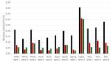
All 9 peptides were subject to ETD at varying supplemental activation energies in the mass spectrometer. The peaks in each spectrum were assigned and any neutral loss peaks plotted against activation energy. Of the peptides studied, only peptide 9 showed any ambiguous fragments (c8-80). The c8-80 fragment of peptide 9 has identical mass to the c8 fragment from peptide 8. Figure 1 shows a of plot of peak intensity against activation energy for the c8 and c8-80 fragments from peptide 9. There is no loss of 80 Da until a supplemental activation energy of 12.5%, at which point the peak intensity rapidly increases up to an activation energy of 25%.
Figure 1. Effect of supplemental activation on the abundance of the c8 and c8-80 fragments of triply-charged peptide 9.
Figure 2 shows the saETD mass spectra obtained for peptide 9 at supplemental activations of 0%, 15% and 25% respectively. The c8-80 fragment is not present at activation energy 0%, however it is apparent when this is increased to 15% and its abundance further increases at 25%. Again you can see from the fragmentation patterns that the number of fragments increases with increasing activation energy. This shows that when considering optimum activation energy, we should not make it too low as we will not see efficient fragmentation.
From the LTQ Orbitrap Velos ETD results, we determined that when carrying out the FAIMS experiment we would use a maximum supplemental activation of 7.5%, as at this activation energy for all of the peptides we do not see any loss peaks. This then allowed us to look at the FAIMS data and analyse any neutral loss peaks knowing that they were solely due to the FAIMS and not saETD. The dispersion voltage was varied because it may cause low levels of collisional activation through collisions with the carrier gas. By reducing the DV, the ions path are slowed reducing the energy transferred.
The FAIMS ETD spectra were analysed for the presence of ambiguous fragments, e.g., neutral loss peaks. The previously identified c8-80 fragment from peptide 9 was not present in the FAIMS data at any of the 3 dispersion voltages, e.g, see Figure 3. No ambiguous fragments were observed in any of the FAIMS data. This result informs us that the FAIMS device does not promote the loss of the phosphate and that in this is example it is solely down to the supplemental activation. This knowledge means that we can confidently carry out experiments using the FAIMS and accurately identify the phosphorylated residues of co-eluting isobaric phospho-peptides.
Figure 3. FAIMS ETD mass spectrum triply charged peptide 9 (APLpSFLGSLPKSYVK) at dispersion voltage of 5000.
Conclusion
From this work it can be concluded that the optimum supplemental activation to carry out ETD is at 7.5, to give enough energy so that there is significant fragmentation, but to inhibit the loss or transfer of the phosphate group. Our results suggest that if the FAIMS ETD is carried out at with a supplemental activation of 7.5%, no fragments resulting from the loss or transfer of the phosphate group are observed. This is advantageous as it will increase the reliability of the phosphopeptide analysis as there will be accurate assignment of the phosphorylated residue.
This experiment was carried out by varying the supplemental activation and the dispersion voltages, however there are many other variables that can be altered in the FAIMS device. Further possible work could be carried out, for example varying the electrode temperature in the FAIMS or altering the helium content of the carrier gas.
Acknowledgements
H.J.Cooper, A.J.Creese, The Mass Spectrometry Lab at The University of Birmingham and the British Mass Spectrometry Society
References
[1] Edelson-Averbukh, M., et al., Gas-Phase Intramolecular Phosphate Shift in Phosphotyrosine-Containing Peptide Monoanions. Analytical Chemistry, 2009. 81(11): p. 4369-4381.
[2] Viglietto, G., et al., Cytoplasmic relocalization and inhibition of the cyclin-dependent kinase inhibitor p27(Kip1) by PKB/Akt-mediated phosphorylation in breast cancer. Nature Medicine, 2002. 8(10): p. 1136-1144.
[3] Syka, J.E.P., et al., Peptide and protein sequence analysis by electron transfer dissociation mass spectrometry. Proceedings of the National Academy of Sciences of the United States of America, 2004. 101(26): p. 9528-9533.
[4] Zubarev, R.A., N.L. Kelleher, and F.W. McLafferty, Electron capture dissociation of multiply charged protein cations. A nonergodic process. Journal of the American Chemical Society, 1998. 120(13): p. 3265-3266.
[5] Sweet, S.M.M., et al., Large Scale Localization of Protein Phosphorylation by Use of Electron Capture Dissociation Mass Spectrometry. Molecular & Cellular Proteomics, 2009. 8(5): p. 904-912.
[6] Creese, A.J. and H.J. Cooper, The effect of phosphorylation on the electron capture dissociation of peptide ions. Journal of the American Society for Mass Spectrometry, 2008. 19(9): p. 1263-1274.
[7] Coon, J.J., et al., Electron transfer dissociation of peptide anions. Journal of the American Society for Mass Spectrometry, 2005. 16(6): p. 880-882.
[8] Coon, J.J., et al., Activated-Ion Electron Transfer Dissociation Improves the Ability of Electron Transfer Dissociation to Identify Peptides in a Complex Mixture. Analytical Chemistry, 2010. 82(24): p. 10068-10074.
[9] Coon, J.J., et al., Supplemental activation method for high-efficiency electron-transfer dissociation of doubly protonated peptide precursors. Analytical Chemistry, 2007. 79(2): p. 477-485.
[10] Xuan, Y., et al., High-field asymmetric waveform ion mobility spectrometry (FAIMS) coupled with high-resolution electron transfer dissociation mass spectrometry for the analysis of isobaric phosphopeptides. Rapid Communications in Mass Spectrometry, 2009. 23(13): p. 1963-1969.
[11] Guevremont, R., High-field asymmetric waveform ion mobility spectrometry: A new tool for mass spectrometry. Journal of Chromatography A, 2004. 1058(1-2): p. 3-19.
[12] Palumbo, A.M. and G.E. Reid, Evaluation of Gas-Phase Rearrangement and Competing Fragmentation Reactions on Protein Phosphorylation Site Assignment Using Collision Induced Dissociation-MS/MS and MS3. Analytical Chemistry, 2008. 80(24): p. 9735-9747.
ILM 51.5 July 2026


-(1).jpg)
.jpg)


.jpg)