-
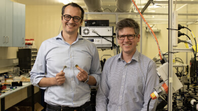 Pictured (left) Alessandro Tuniz and Boris Kuhlmey
Research News
Superlensing Method beats Object Distortion
Oct 18 2023
In attempts to break through the physical limits of observing objects using traditional optical methods, Physicists at the University of Sydney have shown a new pathway to achieve superlensing with minimal losses, breaking through the diffraction limit* by a factor of nearly four times. The key to their success was to remove the super lens altogether.
The work has possibilities for improved super-resolution microscopy, for example in fields as varied as cancer diagnostics, medical imaging, or archaeology and forensics, the researchers said.
Lead author Dr Alessandro Tuniz from the School of Physics and University of Sydney Nano Institute, said: “We have now developed a practical way to implement superlensing, without a super lens. To do this, we placed our light probe far away from the object and collected both high and low resolution information. By measuring further away, the probe doesn’t interfere with the high resolution data, a feature of previous methods.”
Previous attempts have tried to make super lenses using novel materials. However, most materials absorb too much light, such as low resolution data, to make the super lens useful.
Dr Tuniz continued: “We overcome this by performing the superlens operation as a post-processing step on a computer, after the measurement itself. This produces a ‘truthful’ image of the object through the selective amplification of evanescent, or vanishing, light waves.
Co-author, Associate Professor Boris Kuhlmey, also from the School of Physics and Sydney Nano, said: “Our method could be applied to determine moisture content in leaves with greater resolution, or be useful in advanced microfabrication techniques, such as non-destructive assessment of microchip integrity. The method could even be used to reveal hidden layers in artwork, perhaps proving useful in uncovering art forgery or hidden works.”
“By moving our probe further away we can maintain the integrity of the high-resolution information and use a post-observation technique to filter out the low-resolution data.”
The research was done using light at terahertz frequency at millimetre wavelength, in the region of the spectrum between visible and microwave.
Associate Professor Kuhlmey said: “This is a very difficult frequency range to work with, but a very interesting one, because at this range we could obtain important information about biological samples, such as protein structure, hydration dynamics, or for use in cancer imaging.”
Dr Tuniz concluded: “This technique is a first step in allowing high-resolution images while staying at a safe distance from the object without distorting what you see.
“Our technique could be used at other frequency ranges. We expect anyone performing high-resolution optical microscopy will find this technique of interest.”
*The diffraction limit is determined by the fact that light manifests as a wave. It means a focused image can never be smaller than half the wavelength of light used to observe an object.
The research is published in Nature Communications.
More information online

-with-research-lead-Dr-Sandro-Ataide-and-first-author-Rezwan-Siddiquee-(right)---C.-Photo-Fiona-Wolf-USYD.jpg)


Digital Edition
ILM 49.5 July
July 2024
Chromatography Articles - Understanding PFAS: Analysis and Implications Mass Spectrometry & Spectroscopy Articles - MS detection of Alzheimer’s blood-based biomarkers LIMS - Essent...
View all digital editions
24_06.jpg)


Events
Jul 28 2024 San Diego, CA USA
Jul 30 2024 Jakarta, Indonesia
Jul 31 2024 Chengdu, China
ACS National Meeting - Fall 2024
Aug 18 2024 Denver, CO, USA
Aug 25 2024 Copenhagen, Denmark


