Electron
Published over 2 years ago. See the latest and most current information on Electron.
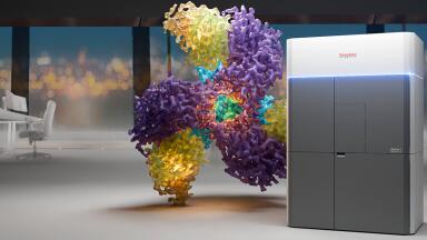
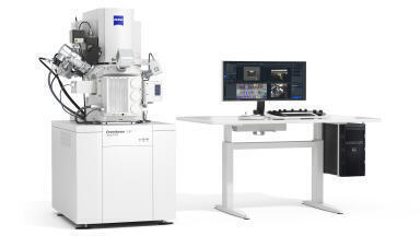
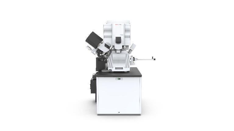
The Thermo Scientific Hydra Bio Plasma-Focused Ion Beam (Plasma-FIB) is a cutting-edge instrument designed to streamline workflows for cell biologists engaged in volume electron microscopy, whether it involves cryo or resin-embedded samples.
Built upon the industry-standard Thermo Scientific Helios Hydra DualBeam platform, the Hydra Bio Plasma-FIB stands as a versatile, multi-application solution catering to volume electron microscopy and sample preparation in the cryo-electron tomography workflow. From studying tissues to delving into proteins, this instrument acts as a bridge, facilitating the transition from cryo-EM to room temperature analysis and vice versa. By offering this simplicity, it empowers cell biologists to channel more of their valuable time and effort into advancing intricate research projects.
With the Hydra Bio Plasma-FIB, researchers can explore a wide array of microscopy techniques, including FIB-SEM serial sectioning (both at room temperature and in cryogenic conditions), array tomography, and cryo-electron tomography. This flexibility empowers biomedical researchers to unravel the three-dimensional intricacies of cells, tissues, and small organisms at a remarkable nanometer resolution.
Users can seamlessly switch between four distinct ion species (Xe, Ar, O, and N) to optimise their approach according to the specific demands of each sample.
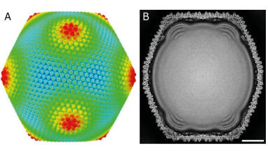
By employing the cryogenic mode, researchers can delve into cellular architecture in its native state, achieving exceptional contrast. This approach enables the exploration of sub-cellular intricacies in high-pressure-frozen and plunge-frozen samples, eliminating the need for staining.
The room temperature mode allows cell biologists can now gain insights into large sample areas and visualise areas of interest with the Spin Mill Bio Method. This innovative technique generates large area planar-milling results similar in size to microtome slicing, but with slice thicknesses as minimal as 5 nm. It facilitates the preparation of clean, smooth surfaces, which are then utilised for locating regions of interest and subsequent imaging in both 2D and 3D.
More information online
ILM 51.5 July 2026


-(1).jpg)
.jpg)