News
Published over 4 years ago. See the latest and most current information on News.
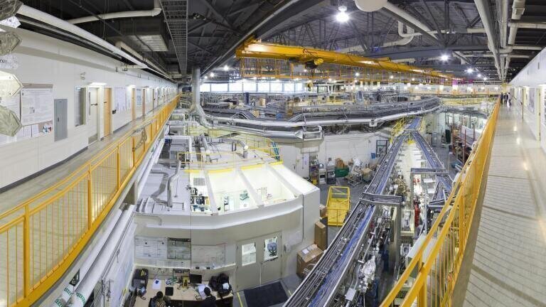
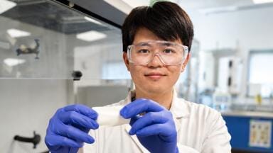
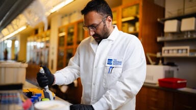
Working towards development of an alternative to the use of bone grafts for repairing diseased or damaged bone, researchers at McGill University have used the Canadian Light Source (CLS) at the University of Saskatchewan to advance a novel method for growing synthetic bone tissue.
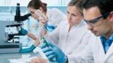
Bone tissue engineering is focused on growing bone cells in scaffolds in the lab and transferring these structures into the damaged area inside the body. The material used for the scaffold needs to have an interconnected network of small and large pores to enable nutrients and cells to spread into the damaged area and help generate new bone tissue. The McGill team took the approach of modifying the internal structure of Graphene oxide an ultrathin, extra strong compound that is being used increasingly in electronics, optics, chemistry, energy storage and biology. One of its unique properties is that when stem cells are placed on it, they tend to transform into bone-generating cells called osteoblasts.
The multidisciplinary group – comprising researchers from McGill’s Departments of Mining and Materials Engineering, Electrical Engineering, and Dentistry – found that adding an emulsion of oil and water to the graphene oxide, then freezing it at two different temperatures, yielded two different sizes of pores throughout the material.
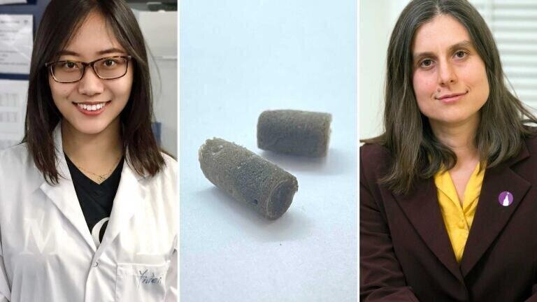
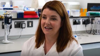
Professor Marta Cerruti said that when they “seeded” the now-porous scaffolding with stem cells from mouse bone marrow, the cells multiplied and spread inside the network of pores, a promising sign the new approach could eventually be used to regenerate bone tissue in humans.
“We showed that the scaffolds are completely biocompatible, that the cells are happy when you put them in there, and that they’re able to penetrate all through the scaffold and colonise the whole scaffold,” she stated.
The researchers used the BMIT-BM beamline at the CLS to visualise the different sized pores inside the scaffolding as well as the growth and spread of the cells. Lead researcher Yiwen Chen, a PhD student working under Cerruti, said their work would not have been possible without the synchrotron because the low density of graphene oxide means it absorbs only a very small amount of light.
“To our knowledge, this is the first time that people have used synchrotron light to see the structure of graphene oxide scaffolds,” said Chen.
Although widespread clinical application of this approach may still be many years away, Cerruti thinks their work could enable other researchers to learn more about how stem cells morph into bone cells.
“Maybe this will lead to a better understanding of the biology of bones that we wouldn't understand otherwise,” she said. “Perhaps in the shorter term we can use the methods in the lab to better understand bone and perhaps develop new drugs.”
More information online
ILM 51.5 July 2026


.jpg)


-(1).jpg)


.jpg)
.jpg)

