
News
Published over 16 years ago. See the latest and most current information on News.
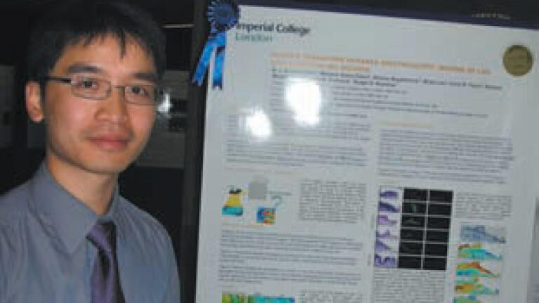
The annual Hounsfield memorial lecture was instituted by the Imperial College London Imaging Sciences Centre to recognise the contribution of Sir Godfrey Hounsfield in the development of medical imaging systems. The lecture has been designed as an opportunity for a world leading researcher in imaging science to review the latest developments in imaging science. This is supported by a range of posters produced by
imaging related research groups in Imperial which demonstrate the scope and depth of research going on at the College.
The fifth lecture in the series was given on 8th June 2009 by Professor James Fujimoto from the Department of Electrical Engineering and Computer Science and Research Laboratory of Electronics, Massachusetts
Institute of Technology. His talk was entitled ‘Biomedical Imaging and Optical Biopsy with Optical Coherence Tomography (OCT)’.
With an audience of over two hundred, Sir Roy Anderson, Rector of Imperial College London, presided over the meeting. He introduced Professor Fujimoto as a man passionate about teaching, a model advocate of interdisciplinary collaboration and a man who has the motivation to make a difference in the world. His contributions in biomedical imaging are an excellent example of him achieving his goal.
Professor Fujimoto covered a broad range of topics giving a history dating back to the early 90s. He charted ups and downs when the technique nearly died through lack of commercial opportunity. However, by 2006 there were over 6,000 systems being used in the ophthalmic field from Carl Zeiss alone with their Stratus system. Retinal imaging, identification of glaucoma and macular degeneration are all now well established applications for OCT.
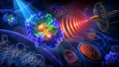
OCT has emerged as an imaging modality which can generate high resolution, cross-sectional and three dimensional images of microstructure in biological systems. Imaging is performed by measuring the echo time delay of light backscattering in tissue. OCT can perform "optical biopsy" enabling the in situ, real time visualisation of tissue pathology. OCT utilises advances in photonics and fibre optics such as femtosecond
broadband lasers, high speed wavelength swept lasers and line scan camera technologies. Three dimensional volumetric imaging with extremely high voxel density is now possible, enabling microstructure and pathology to be visualised and rendered in a manner analogous to magnetic resonance imaging. OCT is rapidly becoming a clinical standard in ophthalmology, where it can image retinal pathology with unprecedented
resolutions. OCT is also being developed for other applications ranging from cancer detection in endoscopy to intravascular imaging in cardiology.
It was clear that the technology is developing at a fast rate as both software and hardware improvements coupled to laser developments have increased sensitivity of measurement while greatly reducing the time taken for the measurement. At this time, OCT is being used as a fast diagnosis tool based mainly on the observations of the user, e.g. the ophthalmologist. Looking to the future, the use of pattern recognition software may well be applied as it has started to become accepted in the field of digital pathology.
Screening and early stage diagnosis of cancers is another area of growth for the use of OCT. Work using an endoscopic probe has been reported where OCT may prove helpful in the early diagnosis of Barrett’s esophagus, damage of the gullet (foodpipe) where refluxing acid from the stomach can damage cells. This in turn may lead to esophageal cancer, the UK’s 9th most common cancer with around 7800 cases being diagnosed each year.
Not all applications are biomedical in nature. Professor Fujimoto demonstrated examples where OCT has been used in applications of art conservation. As the technique enables imaging of sections, it may be applied to the different processes of a painting. For example, the surface is likely to be a varnish. Below this will be the paints/pigments and then below that may be the original charcoal sketches of the artist before the painting proper commenced. An example of a 16th century painting was used to show the power of OCT once again.
The success of OCT may also be measured through observing the number of papers now being published in the field. The last 2-3 years has seen an explosion in publications with over 7,000 currently reported with around half being ophthalmic in origin. To put this into perspective with other microscopies, OCT is chasing after confocal microscopy (29,000). While growth will continue in medical areas, new applications will
develop in areas such as metrology and other areas of imaging below a surface.
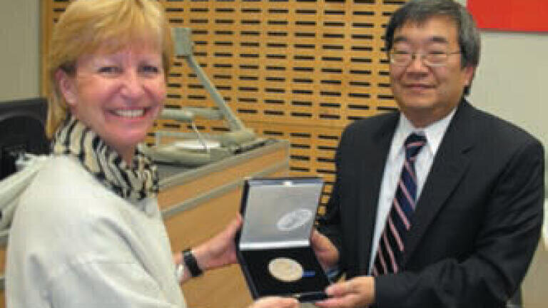
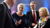
In closing the presentation and proposing the vote of thanks, Professor Maggie Dallman, Principal of the Faculty of Natural Sciences at Imperial, presented Professor Fujimoto with the 2009 Hounsfield medal (pictured below). The 2009 Hounsfield lecture was sponsored by CORDA, the pioneering charity dedicated to preventing heart diseases and stroke, by Michelson Diagnostics and the Clinical Imaging Centre.
ILM Guide 2026/27


.jpg)
.jpg)


.jpg)

2.jpg)
