Microscopy & microtechniques
Published over 5 years ago. See the latest and most current information on Microscopy & microtechniques.
Principal Beamline Scientist Maria Harkiolaki and Support Scientist Chidinma Okolo, explain how two powerful microscopy methods come together to deliver complementary high resolution cryo-imaging of cell structure and compositions at near-physiological conditions.
The biological cryo-imaging beamline B24 at the UK synchrotron Diamond Light Source (https://www.diamond.ac.uk/Instruments/Biological-Cryo-Imaging/B24.html) has developed a high resolution 3D cryo-imaging platform that delivers detailed 3D X-ray and visible light fluorescence imaging of cells and cell populations at near-physiological conditions. The platform has been presented in a recent publication [1] in which, it was used in the study of reovirus vesicle egress in mammalian cells and allowed the tracking of viral capsids within endosomal vesicles across the cytoplasm in 3D in the early hours post infection.
The success of biological research depends on understanding a living system in depth from its basic chemical composition to the structures that make up cells and the interactions within tissues and organisms. Microscopy is fundamental in this process allowing us to directly document the complex architecture of life, but as all research tools, it stands restricted by its subject matter. To see the macroscopic means that the observer gains a top level view of a system but misses the fine details, whereas when we look into the microscopic, context is often sacrificed. For example a CAT scan with a wide field of view will document whole tissue arrangements within an organism but it takes an electron microscope with a field of view of just a few microns to visualise the fine structures within individual cells from that same organism. And for each and every step we take towards ever increasing imaging resolution and detail capture, sacrifices need to be made. Cells need to be extracted from their natural environment, modifications need to be made to highlight them and their very chemical composition needs to be altered to immobilise them - an image can only be as clear as sample movement and vibrations within allow it to be, therefore, becomes imperative when we seek to gain information on cellular structures and fine chemical localisation to engage microscopy techniques with variable resolutions while retaining as much as possible physiological features and relative sizes. Moreover, when progressing the scales of image resolution, we ideally move across methods that overlap in their capability boundaries (resolution increases gradually as field of view collapses) allowing the productive overlap of data from one scale to the next with high resolution imaging benefiting from the wider context supplied by less powerful techniques.
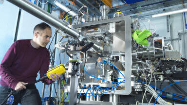
The correlative cryo-imaging beamline B24 at the UK synchrotron Diamond Light Source endeavours to address all of the above and deliver directly correlatable 3D imaging data across scales and contrasting schemes on samples at near-physiological state. The imaging platform at B24 comprises of a novel super resolution fluorescence structured illumination microscope (cryoSIM) and soft X-ray tomography end station (cryoSXT) and allows users to visualise biological samples such as cells and tissue slices.
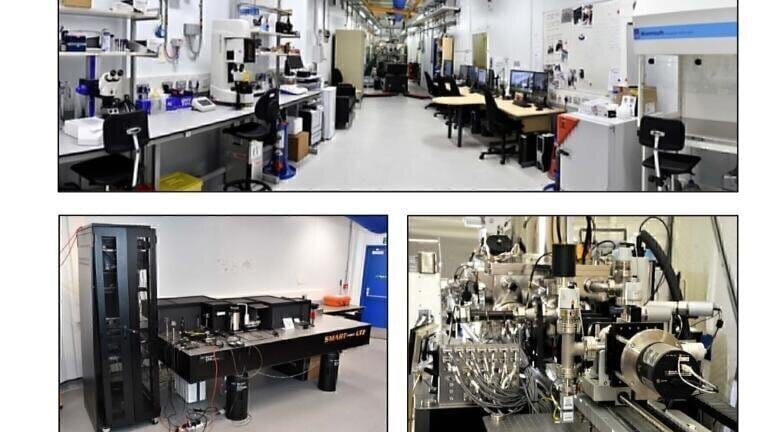
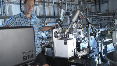
Diamond Light Source beamline B24 main cabin (top panel); the cryoSIM (cryogenic temperature-Structured Illumination Microscope) (bottom left) and cryo-SXT (cryogenic temperature-Soft X-ray Tomography) microscope (bottom right).
It offers 3D cellular correlative imaging at cryogenic temperatures and features (a) 3D data capture, (b) same sample fluorescence and X-ray imaging, (c) minimal sample processing requirements, (d) image resolution to 25nm, (e) rapid data collection and (f) easy to follow access procedures through peer-review processes (for academics). In addition, the beamline also provides access and support on accessory equipment to allow sample preparation and evaluation as well as data processing, analyses and correlation.
Biological imaging across scales and B24 remit
Suitability and requirements for use of the B24 facility.
Current requirements for biological imaging
The requirements for biological/biomedical imaging at the cellular level continue to increase and become more complex as we probe further into biological systems. Information should ideally be captured in 3D, to the highest resolution possible and give us clear views into the architecture and composition of cells and tissue without losing fidelity of information through artefacts induced because of harsh sample preparation procedures or microscopy requirements. At beamline B24, this is achieved via cryo-preservation where cells grown adherent or deposited on grid sample holders (3mm gold wafers coated with a perforated carbon support film) are plunge frozen in liquid nitrogen and then kept cold throughout transfer and exposure to light. This ensures that the sample not only remains perfectly static during imaging but also sustains little or no damage during exposure to the harsh illumination conditions required for high resolution imaging. Moreover, as chemical fixation or embedding are not required, no chemical alterations have been induced within the sample and therefore the risk of misinterpretations is minimised.
Mapped 3mm sample grid carrying human HFF cells (the outline of cells can be seen in the square inset). The panel on the left shows tweezers used for transferring grids and two gold grids on a film-covered surface for size.
Other necessary pre-requisites for successful biological imaging in general are reproducibility, accessibility and ease of use. New technology should deliver reliably, at all times, made available as soon as possible, and have clear access routes. At beamline B24, the correlative cryo-imaging platform has been put through it’s paces for the past year and research groups working on a number of areas from cancer biology, infection and pathogen clearance to bacteria, algae and archaea biology have benefitted from the technology in this time while the beamline team has focused on streamlining user support, accessibility, sample preparation and mapping as well as data collection and processing. Data collection at B24 typically takes 3-5 days per project (time on both cryoSIM and cryoSXT) and results in many Gigabytes of 3D data (10-30 SIM data sets, 1-3 X-ray tomograms per SIM data set) of areas of 100s of microns in and across cells.
Slice of 3D cryoSXT data from human U2OS cells showing the level of detail that can be visualised with soft X-ray tomography and also the organelles and features prominent in a cryo-preserved mammalian cell at near-physiological conditions.
Slice of 3D cryoSXT data (left) showing an area of a cell infected with reovirus (the nucleus and cytoplasm are indicated by a blue and green arrow respectively; the interface of this cell with its neighbour is outlined with orange arrows). CryoSIM data (also 3D) is superimposed (right) to delineate organelles carrying viral load in the perinuclear area two hours post infection. This figure clearly shows the benefits of combining a high-resolution imaging techniques such as SXT (nanometer resolution of cell ultrastructure; organelles and other substructures clearly delineated) with 3D super resolution fluorescence SIM (localisation of particles of interest, reovirus in this case, in 3D). A subset of multivesicular bodies identified near the nucleus in SXT can now be identified with SIM as carriers of viral load.
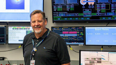
B24 currently has a core team of five scientists all working towards development and automation of the methods offered. They are two biologists, one chemist and two physicists and their expertise range from molecular and cell biology to cryogenic sample handling, beamline instrumentation and software development.
The current B24 core team. Next to the cryoSXT microscope is Dr M Harkiolaki (outer left),
Dr C Okolo (middle left), Mr T Fish (middle right), Dr I Kounatidis (outer right). A new member
Dr M Koronfel has recently joined B24.
The B24 group collecting and evaluating X-ray.
They are supported by beamline technicians, engineers and designers, as well as a host of other support teams within Diamond Light Source from building and facility maintenance to IT and software support and development. All company departments are to a great degree interconnected and provide rapid and relevant support to the beamlines though out the year. An enthusiastic and professional User Office team provides a handy interface between the technology and innovation teams and the user community that engages with the beamlines and use the equipment available.
Development of new techniques transcends the remit of a single organisation and greatly benefits from cross-institutional collaborations. At B24, a productive collaboration with Micron at the Biochemistry department of the University of Oxford in the UK has resulted in the development and delivery of the cryoSIM. This endeavour has in turn seen the support of microscopy and scientific software experts from around the world and continues to develop as a collaborative project.
Software development at B24 is driven by efforts of the core team in close contact with the scientific software, motion and controls group at Diamond Light source but extends beyond the confines of the home institute and involves collaborations with software scientist across the globe. Such a collaboration with Dr P. Paul-Gilloteaux at the University of Nantes in France (the developer of 3D correlation software eC-CLEM) involves the tailored development of bespoke 3D image registration software specifically for SIM and SXT which is currently underway.
Beyond the microscopy and scientific software experts however, it is primarily the researchers themselves that drive and guide developments in imaging technology. Beamline B24 has been privileged to work with enthusiastic and committed research scientists that engaged with the methods, fed back to the design process and drove automation development. As a result, the correlative imaging platform at B24 can now deliver an international first in user-friendly, high-resolution, 3D correlative cryo-imaging. As Dr Stephen Graham, a Sir Henry Dale Fellow and University Lecturer at the Division of Virology of the University of Cambridge commentedz, “The facilities at B24 give us unique tools to probe virus infection in cells. By correlating super-res light cryo-microscopy with soft X-ray cryo-tomography we can understand exactly what stage of infection each cell is at and can probe the structural rearrangements that viruses induce in the host cells that they exploit. The fixation-free and stain-free workflow plus the huge depth of field available using cryoSXT is especially advantageous for us as it allows us to capture rare events within the cell, like catching viruses in the act as they cross the nuclear membrane.”
Dr. S Graham and his collaborator Dr C Crump collecting data on HSV1 infected cells at beamline B24
Dr Gianlucca Tozzi, a reader in Bioengineering and Director of the Zeiss Global Centre at the University of Portsmouth in the UK has used the B24 imaging platform in his studies on biological tissues and biomaterials for tissue engineering. He said: “Working at B24 has been a great experience. Support in all technical aspects has been brilliant and there is a real will to push further development of the technology in addressing complex imaging modalities, which are increasingly needed to unravel biological mechanisms. I am looking forward to continuing this excellent collaboration”
This novel imaging approach is particularly relevant to the field of cancer biology. As Professor Peter Saddler from the Chemistry Department at Warwick University in the UK highlighted: “B24 has added new dimensions to our work on elucidating the mechanisms of action of novel anticancer drugs which can combat resistance to treatment, in particular prodrugs which are activated specifically in cancer cells by visible light. Correlative super-resolution fluorescence microscopy and 3D soft X-ray tomography allows us to monitor both the localisation of fluorescent drugs and drug-induced subcellular changes to organelles, such as mitochondria and endosomes, in cryopreserved cancer cells close to their native state.”
Significantly, industry also acts as a driving force for development at the beamline. As Dr Claire Pizzey, deputy head of Industrial Liaison at Diamond put it: “Industrial engagement is an important aspect of the work of any beamline at Diamond given that up to 10% of operational time can be dedicated to proprietary use for confidential research and development.
Dr G Tozzi’s team collecting data at B24 on the morphology of differentiated stem cells from sheep.
Commercial interest in correlative microscopy at B24 has been growing fast and recent client projects have focused on key areas for the pharmaceutical industry such as novel therapeutics and vaccine development. Of particular value to industrial clients is the beamline’s full service approach, where no prior knowledge is required. The expert team at B24 can support all stages of the project, from experimental design and data collection through to analysis and reporting. Our clients greatly value the scientific insight, gained in a timely manner, which feeds into their own research and development pipelines.“
Often and unexpectedly the major hurdle once a microscopy setup is operational is to let the people that will most benefit from it know that it is now available and how they can take advantage of it in the most time-efficient and productive way. At B24 processes are fully documented on our websites (www.diamond.ac.uk/Users.html and https://www.diamond. ac.uk/Instruments/Biological-Cryo-Imaging/B24.html) and accompanied with protocols and expertise that allow the standardisation of the steps required and remove the element of guesswork from sample preparation and processing as well as data collection and analyses.
At B24, the development of a new imaging platform has filled previously unmet needs of biological imaging by combining the power of cryo-SIM and cryo-SXT microscopes to provide valuable insights into the inner workings of biological material. One method allows us to map the location of chemical signatures and the other gives us ultrastructural information and cellular content, literally a CT scan of a cell with highlighted areas of interest. Once together the researcher has both a 3D map of a cell, and the specific location within it of molecules of interest. This innovative and largely user-friendly microscopy setup at beamline B24 perfectly demonstrates what can be achieved by multidisciplinary collaboration and is now available to the wider research community.
Further information from [email protected]
1. Kounatidis, I. et al. 3D Correlative Cryo-Structured Illumination Fluorescence and Soft X-ray Microscopy Elucidates Reovirus Intracellular Release Pathway. Cell 182, 1–16 (2020).
ILM 51.5 July 2026


-(1).jpg)
.jpg)
.jpg)

.jpg)