Microscopy & microtechniques
Published over 4 years ago. See the latest and most current information on Microscopy & microtechniques.
Everything we do from moving a finger to thinking and memory formation, triggers a signalling cascade in the brain and body. Neurotransmitters (NTs) are released from the synapse - the nerve terminal between neurons where information is passed from one neuron to the next - into the synaptic cleft, the 20-30 nm space between neurons, and onto the beginning of the neighbouring cell which is called the postsynapse (Figure 2).
If movement is involved, eventually the signal from the brain will reach a designated muscle cell instructing it to contract which could lead, for example, to the movement of a finger. Information about a new position of the finger will be immediately sent back to the brain from receptor neurons to check if intended results are achieved or if any correction is required.
If we want to understand how the brain and nervous system is affected by diseases, injury, or mental health conditions, its fine structure and function must be studied. High resolution microscopy techniques play a crucial role in this quest; they are important for visualising the fine details needed to develop treatments.
In the past, brain cells were studied after fixation involving dehydration and staining by chemicals, to preserve a snapshot of the grand structure of neurons in a particular state. However, this meant fine details were lost, such as small protein filaments in synapses that provide communication between the cells. These structures, called tethers and connectors, connect one vesicle to another. It is important to investigate small proteins like these because they are often affected in neurodegenerative diseases.
Cryo-ET is a branch of cryo-Electron Microscopy (cryo-EM). The name of the method underlines that samples are studied at very low temperatures, normally at -196°C. At this temperature water, which makes over 90% of content in all living organisms, can exist in amorphous non-crystalline form, also found in comets in deep space.
During cryo-EM sample preparation, the water in the sample is turned into amorphous ice (called vitrification) by rapid plunging of the sample into liquid ethane, which is cooled down by liquid nitrogen to about -172°C. Biological samples are vitrified so rapidly that ice crystal formation is prevented, and structural integrity preserved as if they are still in their natural environment. The samples are said to be preserved in a frozen-hydrated state, since no drying occurs during sample preparation and study, for vitreous ice can be kept in the electron microscope vacuum at temperatures below -156°C.
In cryo-ET, a 3-dimensional (3D) model of a unique sample can be built by combining multiple projections of the sample taken at different angles. Electrons can travel through the sample in the same way as X-rays during medical tomography, but because electrons are charged, electro-magnetic lenses can be used to create a detailed magnified image of a very small object, allowing us to explore the universe of sub-cellular molecular structures (reviewed in [1]).
Moreover, by subjecting cells to various treatments with chemical and physical stimuli, and then taking snapshots after the treatment, one can potentially study tiny changes. This allows understanding of how the cells function, enabling the creation of a dynamic, 4-dimensional (4D) model of cellular processes.
Cryo-ET produces nanometer-scale information in the form of high-resolution (~1–4 nm) 3D views of samples, typically biological macromolecules, and cells. The technique is ideally suited to study thicker cellular specimen in their native cellular context, but optimal sample thickness is below 300 nm.
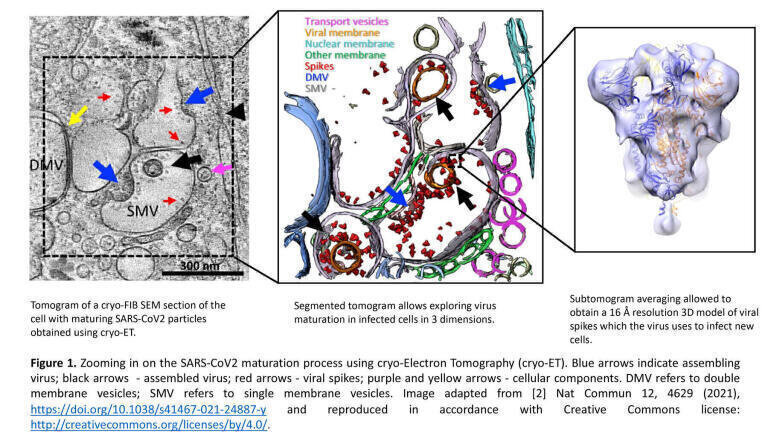
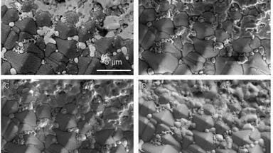
For unlocking details of thicker samples, thinning by Focused Ion Beam milling in a dual beam Scanning Electron Microscope (SEM) has been invented (cryo-FIB SEM). A thin section of the cell can be used for building 3D models using cryo-ET [1]. Application of this technique by scientists at the UK’s electron Bio-Imaging Centre (eBIC) at Diamond Light Source helped to shed light on the life cycle of SARS-CoV-2 at the onset of the global Covid-19 pandemic (Figure 1) [2].
My interest in high resolution microscopy developed after studying the brain for my Master of Sciences (MSc) at the Max Planck Institute of Psychology in Munich, Germany. I wanted to understand the structures in the brain more thoroughly so I investigated which microscopes could achieve a higher resolution and discovered exciting new techniques like cryo-EM, a high-resolution imaging technique awarded the Nobel prize in 2017.
In 2012, I relocated to the Institute of Anatomy in Bern, Switzerland, for my PhD and a short stint as a postdoctoral researcher. The PhD was to study synaptic vesicle exocytosis in synaptosomes (pinched off nerve endings) by cryo-ET - another technique still in its infancy at the time.
I really enjoyed cellular tomography because there are many interesting things inside a cell that catch the eye and make you wonder what they could be. This made me want to investigate all the processes inside the cell but unfortunately time was limited!
Learning the amazing cryo-ET technique, developing, and improving ways to achieve millisecond resolution with an atomizer as originally used by Berriman and Unwin [3], and analysing the tomograms for smallest changes on the synaptic vesicle and the opposing active zone plasma membrane was very intriguing. I also had the opportunity to expand my knowledge in protein biochemistry, cell culture and related sample preparation for cryo-EM processing, as well as learning cryo-FIB. Ultimately, my aim was to unravel ongoing cellular processes studied by cryo-light and electron microscopy.
To delve deeper into neuroscience, I obtained an early career postdoctoral research grant from the Swiss National Science Foundation to join a lab in Copenhagen, Denmark, in 2017. I initially learned electrophysiology and chromaffin cell preparation but then moved on to primary astrocyte and neuron cell culture.
Understanding neurotransmitter release (synaptic vesicle (SV) exocytosis or signal transduction) in healthy neurons - which proteins are involved and what happens during release - are the necessary basics before researching how diseases affect brain functionality.
The synapse contains synaptic vesicles which are filled with signalling molecules. Our research looked at functional aspects of synaptic vesicle exocytosis to understand which proteins are involved and what happens when certain proteins are knocked out through a gene deletion.
Eventually I managed to establish a method to grow neurons on EM grids that also made lots of thin functional synapses also possessing postsynapses (Figure 2). With the right care and conditions growing primary neurons on grids can be straightforward but getting a functional synapse (meaning synapse, cleft and postsynapse) on the grid that is also thin enough for cryo-ET is not easy.
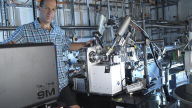
In 2019, I started work as a Senior Support Scientist for Electron Microscopy at eBIC. My previous experience - particularly in cryo-ET - prepared me for this role.
Around 70% of my time is focused on support to UK and worldwide users who send us samples. I help them with experiment set-up such as data acquisition strategies, sample preparation, and advise on what to improve. We also work on other projects as well as our own.
For example, after the Covid-19 pandemic started we dedicated all our resources to help investigate the Covid-19 virus to beat the disease. We investigated the viral spike protein and worked on cellular samples to see what the very first vaccines being developed were doing in the cells.
Single-particle cryo-EM was used to investigate the spike protein isolated from the virus itself to unravel the spike’s structure [4] and cryo-ET for cellular investigation. By knowing how the spike enters the cell and which structure it uses, specific drugs could be developed to interfere with the structure to prevent cell entry [2]. This was complemented by other advanced X-ray imaging and diffraction techniques available to us at Diamond. Combining soft X-ray imaging with cryo-ET and serial Focused Ion Beam scanning electron microscopy allowed a detailed picture of SARS-CoV-2 infection to be described [Mendonca et al [2]]. X-ray crystallography provides high resolution structural data that can be used to accelerate the drug discovery process and is being successfully used at Diamond to understand, for example, how variants of concern escape from natural or vaccine induced immunity [reference – Cell papers by Stuart et al [5]].
The other 30% of my role at eBIC is research time on my own project to better understand the brain by investigating the relationship between brain structure and function. Understanding this in a healthy system can improve how we understand systems affected by disease or mental health conditions to develop treatments. I am assisted by a Year in Industry student working with primary neuron cultures and acquiring tomograms to study small proteins within the synapse.
We employ a range of high-resolution microscopy techniques using the latest technology at eBIC. I find it fascinating to learn as much as I can about the technical details of a microscope. It is great troubleshooting microscope challenges together with the engineers at Diamond, as well as learning how to solve some of the easier problems myself. Recently I found myself inside the box of a Talos Arctica TEM trying to find a specific cable that needed to be unplugged to reset a connection!
Having the motivation to learn about the physical/optical properties of a microscope is useful for a deeper understanding of microscopes and how to operate them.
In my case, I came to my field of research with a general biology background and specialised into neuroscience. I participated in an intensive two-week microscopy course during my biology undergraduate degree, during which time I learnt how to use light microscopes (LMs). I progressed to using a confocal LM during my MSc project, and from there moved to EM during my PhD.
Now I teach EM and neuroscience to a third year Bachelor of Science student. She is very motivated and eager to learn all the details of how to operate the microscope and how to grow neurons.
At Diamond, there are several X-ray facilities which can be used to study the same problem but at different scales, from imaging larger volumes using tomography or coherent imaging or studying the chemistry within cells using spectroscopic techniques. There is a lot of opportunity for collaboration both at Diamond and with research facilities around the campus. Because the working environment is pleasant and flexible, there is plenty of scope to develop and progress in the field of EM, as well as other fields and techniques. To experience these techniques hands on and discover more, there are plenty of career options at Diamond like applying for a Year in Industry, a Summer Placement, or a PhD.
Ultimately, one needs to be curious and motivated to learn new things, alongside a willingness to adapt ideas and opinions. With the right mindset there’s always a role that suits.
1. Turk, M. and Baumeister, W. (2020), The promise and the challenges of cryo-electron tomography. FEBS Lett, 594: 3243-3261, https://doi.org/10.1002/1873-3468.13948
2. Mendonça L, Howe A, Gilchrist JB, Sun D, Knight ML, Zanetti-Domingues LC, Bateman B, Krebs AS, Chen L, Radecke J, Sheng Y, Li VD, Ni T, Kounatidis I, Koronfel MA, Szynkiewicz M, Harkiolaki M, Martin-Fernandez ML, James W, Zhang P. Correlative multi-scale cryo-imaging unveils SARS-CoV-2 assembly and egress. Nat Commun 12, 4629 (2021), https://doi.org/10.1038/s41467-021-24887-y
3. John Berriman, Nigel Unwin, Analysis of transient structures by cryo-microscopy combined with rapid mixing of spray droplets, Ultramicroscopy, Volume 56, Issue 4, 1994, Pages 241-252, ISSN 0304-3991, https://doi.org/10.1016/0304-3991(94)90012-4
4. Dejnirattisai, W., Zhou, D., Ginn, H. M., Duyvesteyn, H. M. E., Supasa, P., Case, J. B., Zhao, Y., Walter, T. S., Mentzer, A. J., Liu, C., Wang, B., Paesen, G. C., Slon-Campos, J., López-Camacho, C., Kafai, N. M., Bailey, A. L., Chen, R. E., Ying, B., Thompson, C., …, Radecke J, … Screaton, G. R. (2021). The antigenic anatomy of SARS-CoV-2 receptor binding domain, Cell, Volume 184, Issue 8, 2021, Pages 2183-2200.e22, ISSN 0092-8674, https://doi.org/10.1016/j.cell.2021.02.032 (https://www.sciencedirect.com/science/article/pii/S009286742100221X)
5. Stuart et al : Reduced neutralization of SARS-CoV-2 B.1.617 by vaccine and convalescent serum DOI: 10.1016/j.cell.2021.06.020 June 2021; The antibody response to SARS-CoV-2 Beta underscores the antigenic distance to other variants DOI: 10.1016/j.chom.2021.11.013 Nov 2021; SARS-CoV-2 Omicron-B.1.1.529 leads to widespread escape from neutralizing antibody responses DOI: 10.1016/j.cell.2021.12.046 Jan 2022
Electron Bio-Imaging Centre (eBIC): https://www.diamond.ac.uk/Instruments/Biological-Cryo-Imaging/eBIC.html
Career options at Diamond Light Source: https://www.diamond.ac.uk/Careers.html
Figure 1 Image adapted from [2] Nat Commun 12, 4629 (2021), https://doi.org/10.1038/s41467-021-24887-y. Reproduced in accordance with the Creative Commons license: http://creativecommons.org/licenses/by/4.0/.
ILM Guide 2026/27

.jpg)


.jpg)


.jpg)