Fluorescence
Published over 3 years ago. See the latest and most current information on Fluorescence.
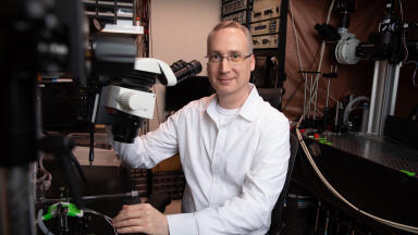
University of Rochester Professor Michael Giacomelli recently received NIH funding for his project, ‘Fluorescence microscopy for evaluation of Mohs surgical margins’. His research objective is to enable real-time evaluation of pathology in skin tissue with an order of magnitude reduction in processing time as compared to frozen sections.
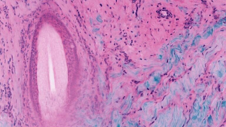
The most common forms of cancer worldwide are basal cell carcinoma and squamous cell carcinoma. Mohs surgery is a widely used technique for the treatment of nonmelanoma skin cancer that obtains extremely low recurrence rates by imaging tissue as it is removed from the body to ensure complete resection. Mohs Micrographic Surgery, also called Mohs Surgery, is a specialised technique to remove non-melanoma skin cancers.
Nonmelanoma skin cancer (NMSC) is primarily diagnosed by histologic analysis of skin biopsy specimens in kerosene sections, which takes days to weeks before a formal diagnosis is made. Two-photon fluorescence microscopy (TPFM) has the potential for point-of-care diagnosis of NMSC and other dermatologic conditions, which could allow diagnosis and treatment at the same site.
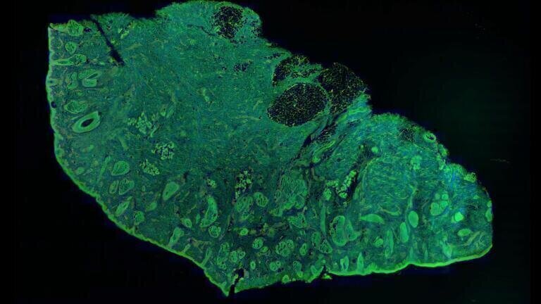
Professor Giacomelli developed a way to image multiple depth slices without frozen section processing‚ by implementing two-photon imaging, widely used in the neurosciences to noninvasively image slices at different depths in living brains. The lab team's research has adapted these fluorescence imaging technologies with rapid tissue labelling and image processing technologies. This enables real-time assessment of pathology in skin tissue. processing time compared to frozen sections has been reduced by an order of magnitude. The data collection time is further reduced using the lower-noised silicon photomultiplier detectors developed by Giacomelli’s group.
2-photon fluorescence microscopy can provide rapid point-of-care diagnosis of non-melanoma skin cancer through real-time imaging of fresh tissue biopsies.
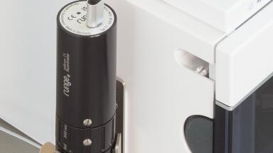
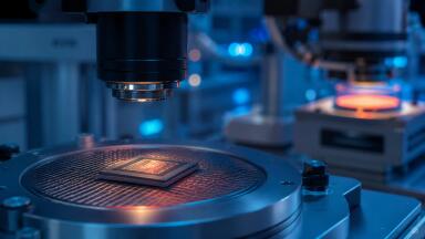
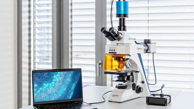
Toptica Photonics’ femtosecond laser system - FemtoFiber ultra 920 - chosen for the next iteration of the Giacomelli lab’s microscope is a possible solution for applications in non-linear microscopy like two-photon excitation of fluorescent proteins and SHG based contrast mechanisms. With an emission wavelength of 920 nm, it provides the highest peak power for imaging with green and yellow fluorescent protein markers (GFP, YFP) commonly used in pathology, neurosciences and other laser-related biophotonic disciplines. Researchers appreciate the usability of the system since it is both, maintenance free and very compact.
More information online
ILM 51.5 July 2026


-(1).jpg)
.jpg)

-(3).jpg)