Microscopy & microtechniques
Published over 12 years ago. See the latest and most current information on Microscopy & microtechniques.
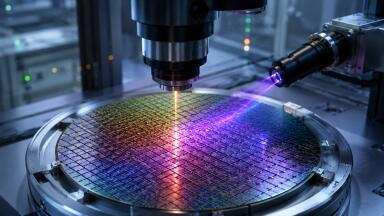
Microscopy is the practice of using a microscope to examine samples and substances that are not visible to the naked eye due to their diminutive size. Its discovery has revolutionised science, and in particular, biology.
There are three main branches of microscopy: optical microscopy, which is divided into many sub-techniques, electron microscopy, and scanning probe microscopy. These are not the only techniques in existence, but they are the best-known and most commonly-used.
Optical Microscopy
This is the most common and well-known type of microscopy, and involves magnifying the image of the object by passing light through or reflecting light off it, and then examining this light through a single or multiple lenses. Traditionally, the image would have to be viewed via the naked eye through a lens; although recent developments in camera technology now mean that it can be captured on a photographic plate or viewed digitally on a computer screen.
There are a wide variety of subdivisions of optical microscopy. These include bright field, dark field, oblique illumination, fluorescence, phase contrast, confocal, deconvolution, differential interference contrast and dispersion staining microscopy, to name a few.
Optical microscopy has several drawbacks; firstly, the technique works at its best only with darker objects, or ones that refract effectively. Secondly, image clarity is often reduced from interfering light outside of the focal plane. And thirdly, the resolution is severely limited by diffraction.
Electron Microscopy
Electron microscopy replaces traditional lenses electromagnets, utilising an electron beam to create an image. This has a far smaller wavelength and as such can improve resolution significantly; up to a factor of 1,000, in fact.
There are two main subdivisions of electron microscopy, which are transmission electron microscopy (TEM) and scanning electron microscopy (SEM), which are comparable to the compound light microscope and the stereo light microscope, respectively.
The various merits of each method, and how they apply in particular to the research of nanocrystals, are explored in more depth in the article Electron Microscopy Reveals Secrets of Nanocrystal-Assembly in Coral Skeletons.
Scanning Probe Microscopy
Scanning probe microscopy differs from other types of microscopy in that rather than “seeing” the image, the instrument “feels” it by probing its surfaces and sending data to a computer, from which the image can be created. There are several types of scanning probe microscopes, all of which are designed to give a more accurate picture of nanoscale structures, surfaces and particles.
Equipment
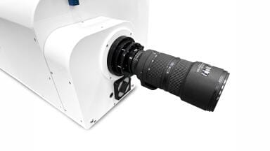
For anyone looking to purchase any of the various types of microscopes mentioned above, see our Microscopy Buyer's Guide. From Electron to Confocal Laser, you'll find the details of all the leading manufactures in the field.
ILM 51.5 July 2026


-(1).jpg)
.jpg)