News
Published over 6 years ago. See the latest and most current information on News.
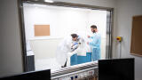
A pre-clinical 3T PET-MR imaging system has been installed by MR Solutions at Western University’s ImPaKT Laboratory in Ontario.
The ImPaKT facility is a one-of-kind facility combining PHAC certified containment level standards (CL2+ and CL3) with advanced in barrier enclosed vivo imaging modalities. These allow researchers to safely develop tools and methods to better understand the progression of infectious diseases, identify efficacious antimicrobial agents, develop diagnostic reagents to characterise hidden reservoirs of pathogens and for the early and accurate detection of infections.
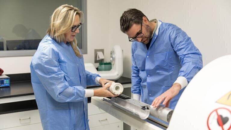
The 3T PET-MR system will be used for anatomical imaging, cell tracking and molecular imaging in virus and pathogen research. Paula Foster, PhD, who will be working in the new laboratory explained that “all kinds of experiments that we were never able to do before with viruses or pathogens and imaging can be carried out. The experiments that we can now do in this laboratory will be entirely new.”
The ImPaKT facility includes MR’s 3T PET/MRI system, the bioluminescence (BLI)-CT scanner, multispectral optoacoustic tomography (MSOT), multiphoton Microscopy, a high resolutions microscope, flow cytometry as well as a GLP PCR clean room, a viral vector core and barrier enclosed animal housing.
ILM 51.5 July 2026


-(1).jpg)
.jpg)


.jpg)