Laboratory products
Published over 3 years ago. See the latest and most current information on Laboratory products.
Transfection methods in mammalian cells are used to introduce or alter genetic material in cells and are often required for larger experimental studies such as stable cell line production, studying gene regulation, mutational analysis, or for mass production of protein.
Transfection methods are usually less than 100% successful; cell type and target gene type can affect success rate. It is therefore important to measure transfection efficiency, which reports the percentage of cells expressing the transfected gene of interest within the cell population.
To enable transfection efficiency to be determined, a reporter gene encoding a product that can be quantified is often attached to the target gene of interest. Reporter genes are typically either fluorescence proteins, enzymes, or substrates that can subsequently be measured enzymatically.
Fluorescence proteins are advantageous as they can be measured in live cells; this approach is typically assessed using microscopy or FACS analysis for single cells. These approaches whilst effective are labour intensive and require expensive specialist equipment.
Multi-mode microplate readers with fluorescence, luminescence, and ultra-violet (UV)-visible absorbance capability such as the VANTAstar® from BMG LABTECH can offer an easy-to-use, cost-effective alternative to measure these assays.
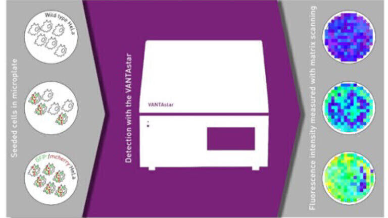
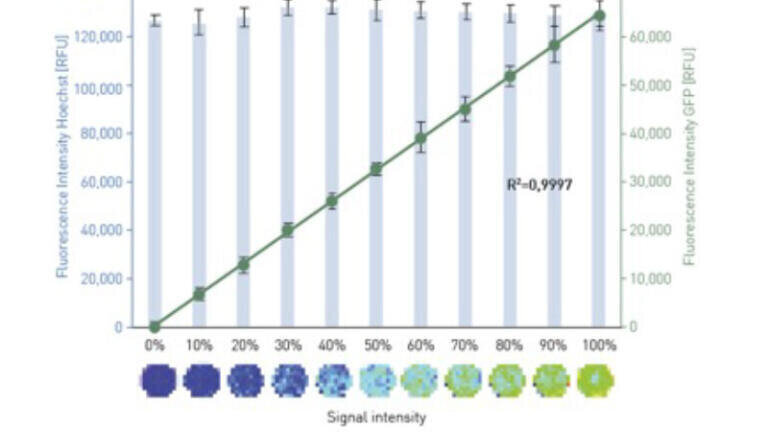
This is demonstrated by quantifying known ratios of wild type (WT)-HeLa cells to GFP expressing HeLa cells. 20,000 cells were plated with different ratios from 0% (WT Hela cells) up to 100% (GFP Hela cells). GFP signal was measured along with total cell number, using Hoechst 33342.
The VANTAstar using a 15 x 15 matrix scan with a diameter of 6 mm, and bottom read detection, was used to capture the fluorescence of both the GFP and Hoechst 33342 signals. Wavelength pre-sets are available in the VANTAstar’s fluorophore library for all common proteins and dyes. The VANTAstar’s LVF Monochromator™ provides full flexibility to measure fluorescence proteins and dyes from the UV to the infra-red (IR) range at industry leading sensitivity limits.
The GFP fluorescence signal shows a linear relationship with the percentage of GFP labelled cells showing an accuracy of r2=0.9997 and a precision of 10.5% CV. The consistent Hoechst 33342 signal confirms the reliability of plating and total cell number.
A multi-mode microplate reader such as the VANTAstar offers an automated alternative to a microscope for the detection of transfection efficiency and other cell-based assays. The flexible LVF Monochromator™ design of the VANTAstar enables users to select the best assay approach without the need to compromise on assay sensitivity. In addition to these fluorescence assays, users can also select different assay readouts in absorbance and luminescence, and can control the environment in the microplate reader, setting temperature, shaking and gas concentrations to maintain live cell samples.
More information online
ILM 51.5 July 2026


.jpg)
.jpg)


-(1).jpg)

.jpg)
.jpg)

.jpg)
.jpg)
-(1).jpg)