Aperio Technologies, Inc has launched TMALab II, the latest release of the company's web-based digital pathology information management system, built specifically for researchers who use Tissue Microarrays (TMAs). With this release, Aperio continues to build upon its commitment to develop digital pathology solutions that meet the evolving needs of pathologists, researchers, and laboratories around the world.
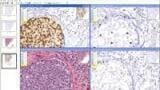
TMALab II software enables researchers to analyze and manage hundreds of small tissue samples simultaneously in a meaningful way to facilitate leading research applications such as new biomarker validation, or discovering and dissecting molecular pathways.
The software is the only web-based product that facilitates multi-user access and server-based image analysis, allowing numerous remote pathologists to collaborate on large TMA projects. Researchers can easily retrieve all stains for any given core, and all cores for any given specimen. Additionally, the software provides links from any TMA core back to the original H&E slide cut from the corresponding donor block.
"Our first generation TMALab product has been successfully assisting scores of TMA researchers to streamline high-throughput tissue analysis in both the bio-pharma and cancer research arenas for many years," stated Dirk Soenksen, CEO of Aperio. "TMALab II enhances the value of our digital pathology solution, sets new standards for ease of integration, and significantly expands data analysis and reporting capabilities for the TMA environment."
Beta versions of the software have been successfully deployed at the National Cancer Institute in Bethesda, MD and the Netherlands Cancer Institute in Amsterdam. TMALab II is currently being used at the National Cancer Institute's Tissue Array Research Program led by Dr. Stephen Hewitt. The initial project involves multiple slides prepared from 32 tissue microarrays. The TMA slides will be scanned using an Aperio ScanScope?, annotated by a pathologist accessing the software via the Internet, and scored using quantitative image analysis algorithms. Additional projects have already been initiated using the platform to examine more than 75 stains on a single TMA.


.jpg)
.jpg)


-(1).jpg)
.jpg)


2.jpg)
