Microscopy & Microtechniques
Complete System for the Visualization of Dynamic Processes at the Cell Membrane
Feb 14 2006
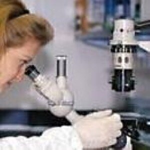
Carl Zeiss presents the Laser TIRF (Total Internal Reflection Fluorescence) Imaging System for cell and molecular biology. The examination of dynamic processes near the membrane of living cells or on the cover slip of cell-free systems has only become possible with the introduction of TIRF. In the past, such a procedure was technically very complex and sensitive to the alignment of the laser beam, i.e. it was accessible only to a few users. Thanks to the modular design of the TIRF imaging system, Carl Zeiss has made the new technology available to a large group of users.
The observation of the dynamic behavior of proteins during transport processes, for example, or of endocytosis and exocytosis on a molecular level requires high spatial and time resolution during imaging. It also necessitates the combination with other observation techniques and incubation possibilities to ensure observation in stable positions over long periods of time.
The Laser TIRF imaging system used with the Axiovert 200 inverted research microscope from Carl Zeiss is the first to offer the quick change of laser lines by means of a fast double filter wheel and the combination of TIRF and transmitted-light contrasting techniques (DIC / brightfield), enabling sequential recording of two image pairs per second with AxioVision software. Carl Zeiss is the only manufacturer to offer the possibility of combining multi-color TIRF, epi-fluorescence and transmitted-light contrasting techniques (DIC) with specimen incubation under laser safety conditions. Incubation of the sample under laser safety conditions is now possible with all stage versions (fixed, heating, mechanical and scanning stages). The special benefits of the scanning stage include higher long-term stability and more precise specimen positioning. For multi-color TIRF, a 100 mW multi-line argon laser with wavelengths of 458, 488 and 514nm is available. Thanks to the optical design, the TIRF effect is implemented for all three wavelengths with a single angle setting. Realignment is not required after the wavelength or the specimen is changed. The three laser lines are ideal for most of the living dyes, e.g. the fluorescent proteins CFP, GFP, YFP and mRFP. A new feature is the TIRF aligning aid for divergence and TIRF angles based on AxioVision software version 4.5 that permits very easy on-screen alignment without compromising laser safety.
Unlike conventional wide-field fluorescence, TIRF offers very high axial resolution as the excitation field penetrates only 100 to 200nm into the specimen. Structures can be excited and therefore visualized only in this volume. Interfering background fluorescence is a thing of the past. The main difference between TIRF and confocal laser scanning microscopy is that the typical thickness of a confocal section measures 400 to 1000nm, while the TIRF method produces a section thickness of approx. 150nm. Therefore, only TIRF permits production of the resolution required at the transition from the specimen to the cover slip (i.e. at the cell membrane) to enable visualization of the dynamic processes of single cells.

Digital Edition
ILM 49.5 July
July 2024
Chromatography Articles - Understanding PFAS: Analysis and Implications Mass Spectrometry & Spectroscopy Articles - MS detection of Alzheimer’s blood-based biomarkers LIMS - Essent...
View all digital editions
24_06.jpg)


Events
Jul 28 2024 San Diego, CA USA
Jul 30 2024 Jakarta, Indonesia
Jul 31 2024 Chengdu, China
ACS National Meeting - Fall 2024
Aug 18 2024 Denver, CO, USA
Aug 25 2024 Copenhagen, Denmark


