Microscopy & Microtechniques
Near Cell Membrane Visualisation
Jun 09 2009
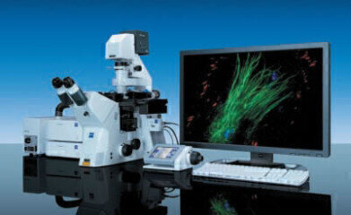
With the introduction of the Laser TIRF 3 microscope system, Carl Zeiss significantly enhances the capability of scientists to visualise near-cell membrane dynamic processes while maintaining optimum specimen incubation conditions. Single molecule dynamic processes in cell-free systems may also be observed and, in combination with other techniques such as Atomic Force Microscopy (AFM), the new microscope provides a complete solution for users in the life sciences, biochemistry, molecular biology and biophysics arenas.
The Laser TIRF 3 maintains Carl Zeiss’ long-standing commitment to system flexibility. A range of incubation options maintains viable conditions for live cell experiments. Together with the Definite Focus module, users can be assured of accurate quantitative data over long time periods. The new laser module may be equipped with up to four solid-state lasers, is AOTF-controlled (Acousto-Optical Tuneable Filter) and may be operated entirely from the AxioVision software interface. The TIRF slider is available in two versions: either manual or fully motorised and software controllable. The motorised version permits a given illumination angle to be set with significantly greater accuracy and speed than other current systems and the reproducible angle setting results in reproducible penetration depths for the light beam. Together with the corrected beam-path and special filter sets, the apochromatically corrected optics of the TIRF slider guarantee maximum image quality.
AOTF control and angle setting are integrated into the ‘Fast Image Acquisition’ module of the AxioVision software, enabling significantly more high resolution images to be acquired within any given timeframe. The new Laser TIRF 3 builds on the attributes of the Laser TIRF, the first microscope to offer the combination of TIRF and transmitted-light contrasting techniques, such as DIC and brightfield, and enable the
sequential recording of image pairs. By selectively exciting cellular fluorophores adsorbed, adhered, or bound to the surface and combining it with conventional epi-fluorescence, researchers can relate surface
effects to internal cellular structures.

Digital Edition
ILM 49.5 July
July 2024
Chromatography Articles - Understanding PFAS: Analysis and Implications Mass Spectrometry & Spectroscopy Articles - MS detection of Alzheimer’s blood-based biomarkers LIMS - Essent...
View all digital editions
24_06.jpg)


Events
Jul 28 2024 San Diego, CA USA
Jul 30 2024 Jakarta, Indonesia
Jul 31 2024 Chengdu, China
ACS National Meeting - Fall 2024
Aug 18 2024 Denver, CO, USA
Aug 25 2024 Copenhagen, Denmark


