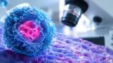
The interiors of cells can now be imaged in three dimensions by laboratory scientists, without causing damage to the cellular structure.
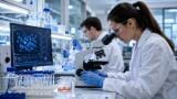
Dr Gerd Schneider and his team have created a single-step solution for imaging cells which does not require them to be dyed, cut or chemically fixed.
The Institute for Soft Matter and Functional Materials researcher has already studied mouse adenocarcinoma cells successfully.
Precision using the new technique is high - down to an accuracy of 30 nm - and achieved by lighting the target object using partially coherent light.
The natural contrast between water and organic matter allows ultrastructures to be visualised by the laboratory scientists at higher contrast ratios than were previously attainable.
Part of the Helmholtz Association of German Research Centres, the institute is also known as the Helmholtz-Zentrum Berlin.
The structure of matter is one of six fields in which the research group is interested, along with energy, health, key technologies, Earth and the environment, and transport and space.