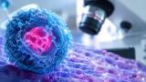
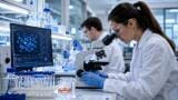
Detecting cancer cells could become much faster with the
latest microscopy technique to be developed at the University of Illinois at Urbana-Champaign.
Scientists at the educational institution have been working with the
latest microscopy technology to create new methods of tissue imaging.
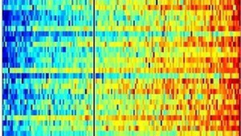
They have developed non-linear interferometric vibrational imaging (NIVI), which works by locking on to the unique vibrational signature of molecules.
By resonating a tissue sample, NIVI can yield a colour map of the cells which highlights potentially cancerous areas in red.
Stephen A Boppart, professor and physician at the institution, says: "We want to make the process of medical diagnostics more quantitative and more rapid."
The University of Illinois at Urbana-Champaign counts astronauts, Olympic medallists, Pulitzer and Nobel Prize winners among its alumni.
Its aims include a pledge to benefit its state, nation and planet by addressing the needs of society through its student programmes and knowledge creation.