Microscopy & microtechniques
Published over 15 years ago. See the latest and most current information on Microscopy & microtechniques.
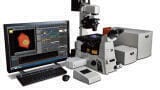
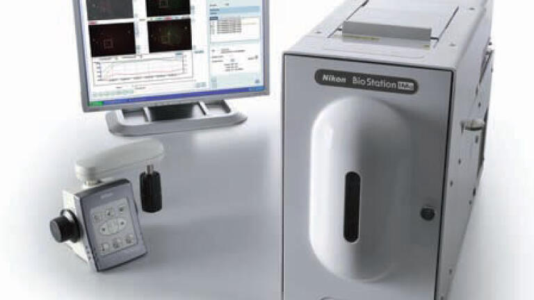
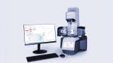
Adding to Nikon’s groundbreaking series of BioStation incubator imaging systems, which offer excellent cell care throughout imaging, the new BioStation IM-Q allows users with minimal microscopy experience to conduct live cell imaging without a steep learning curve. This compact system incorporates a microscope, an incubator and a high sensitivity, cooled quantitative CCD camera integrated into a single package. Providing a stable environment for live cells and advanced phase and fluorescence imaging solutions for simple, long term, cell friendly timelapse data acquisition, the BioStation IM-Q eliminates the need for a darkroom, meaning it can be installed anywhere.
The BioStation IM-Q provides fully motorised control from a PC, allowing users who are not accustomed to operating a microscope or camera to easily conduct timelapse imaging. Integrating cell culture and image capture functions, no complex setup or alignment procedures that conventional timelapse observation systems require, are necessary. Providing thermal and mechanical stability, BioStation IM-Q greatly reduces focus drift, enabling reliable imaging even over long periods.
Two high performance monochrome Nikon Digital Sight camera options are available. The camera’s high sensitivity reduces exposure time, minimising photobleaching and damage to specimens, while increasing throughput for multipoint acquisition. Images at different Z-axis planes can be selected from the captured Z-stack images at each time point and assembled into a seamless movie file – ideal for imaging a specimen
in which the observation point moves along the Z-axis direction, such as with cell division. The streaming function enables rapid motion changes such as cardiomyocyte beats to be captured by high-speed 10-fps imaging at user-defined time intervals. An ergo controller allows X, Y and Z directional movement with an operational feel similar to a microscope.
Two kinds of analysis software are available. Nikon’s proprietary imaging software, NIS-Elements Ar, allows multi-dimensional image capture, image processing, and data management and analysis of up to 6D. CL-Quant software automatically detects and measures the cellular area in unstained, label-free phase contrast images. Unique image processing algorithms provide accurate thresholding of phase contrast images,
enabling non-invasive quantitative analysis of cells. Cell detection accuracy can be improved through a learning function process.
Optional accessories broaden the range of applications.
ILM 51.5 July 2026


-(1).jpg)
.jpg)