News
Published over 6 years ago. See the latest and most current information on News.
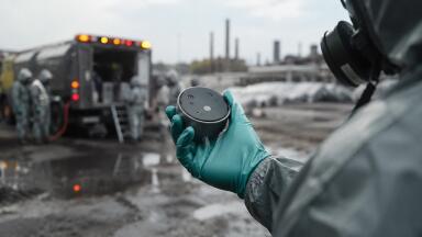
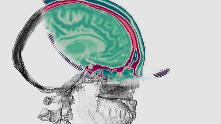
A new computational technique(1) developed by scientists at UCL and Imperial College London could provide doctors with a method for rapid, finely detailed brain imaging using sound waves. They envisage that a small compact device capable of delivering ultrasound would have the potential for portability via ambulance to enable fast assessment of patients prior to arrival at hospital. The researchers are confident the technology will be safe as it uses similar sound intensities as existing ultrasound scanning. With the device designed to be worn like a helmet it also overcomes barriers to bone penetration.
The new approach is of special value in patients investigated for stroke – the second commonest cause of death and commonest cause of adult neurological disability – where rapid, universally applicable, high-fidelity imaging is essential.
Lead author Dr Lluís Guasch, of Imperial’s Department of Earth Science and Engineering, said: “An imaging technique that has already revolutionised one field – seismic imaging – now has the potential to revolutionise another – brain imaging.”
Professor Bryan Williams, Director, NIHR UCL Hospitals Biomedical Research Centre, which partly funded the research, said: “This is an extraordinary and novel development in brain imaging which has huge potential to provide accessible brain imaging in routine clinical practice to evaluate the brain in head trauma, stroke and a variety of brain diseases.
“If this lives up to its promise it will be a major advance. It is also a fabulous illustration of how the collaboration between engineers and clinicians, using methods from another sphere of science, can bring ground-breaking innovation into medical care.”
The computational technique is based on full waveform inversion (FWI), a method that earth scientists use to map the inside of the earth, where seismic data from earthquake detectors (seismometers) are plugged into FWI algorithms that extract 3D images of the Earth’s crust.
In a similar way the new method extracts data from reverberations of sound throughout the skull building a 3D image of the interior.
The researchers developed a helmet lined with an array of acoustic transducers that each sends sound waves through the skull. The ultrasound energy that propagates through the head is recorded and fed via the helmet into a computer. FWI is then used to analyse the reverberations of the sound throughout the skull, constructing a 3D image of the interior.
Dr Guasch said: “This is the first time FWI has been applied to the task of imaging inside a human skull. FWI is normally used in geophysics to map the structure of the Earth, but our collaborative, multidisciplinary team of earth scientists, bioengineers and neurologists are using it to create a safe, cheap and portable method of generating 3D ultrasound images of the human brain.”
Study co-author Professor Parashkev Nachev (UCL Queen Square Institute of Neurology) said: “This is a vivid illustration of the remarkable power of advanced computation in medicine. Combining algorithmic innovation with supercomputing could enable us to retrieve high-resolution images of the brain from safe, relatively simple, well-established physics: the transmission of soundwaves through human tissue.
“The practicalities of MRI will always limit its applicability, especially in the acute setting, where timely intervention has the greatest impact. Neurology has been waiting for a new, universally applicable imaging modality for decades: full-waveform inversion could well be the answer.“
Next, the researchers will build a new prototype for live imaging of normal human brains as the first step to a device that could be evaluated in clinical contexts.
The study was funded by Imperial’s Excellence Fund for Frontier Research, Wellcome, and the UCLH NIHR Biomedical Research Centre.
1) “Full-waveform inversion imaging of the human brain” by Lluís Guasch, Oscar Calderón Agudo, Meng-Xing Tang, Parashkev Nachev and Michael Warner, published 6 March 2020 in npj Digital Medicine.
ILM 51.5 July 2026


-(1).jpg)
.jpg)


.jpg)
.jpg)
.jpg)