.jpg)
News
Published over 3 years ago. See the latest and most current information on News.
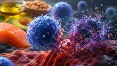
A study led by researchers at Queen Mary University of London (QMUL) has shown that Melanoma skin cancer cells can radically rewire their internal power systems to drive their spread to other parts of the body. The investigation also suggests that reversing this change can make tumour cells less invasive and identified a key molecule that orchestrates this process – knowledge that could lay the foundations for new therapeutic strategies to halt the spread of cancer.
Cancer cells’ ability to break away from the original tumour and spread to other parts of the body (metastasis) seeds secondary tumours that grow in other organs, ultimately causing most cancer deaths.
“We’re still not targeting the secondary disease enough in the clinic and I think we need to change this,” said Professor Victoria Sanz-Moreno, lead author of the new study based in Queen Mary’s Barts Cancer Institute. “In our lab, we want to understand: what are the characteristics of cells that are able to metastasise? What are their weaknesses? And how do we target them?”
Using a three dimensional model system, the researchers observed that metastasising tumour cells adopt a style of movement known as rounded-amoeboid migration, where cells maintain a loose connection to their surroundings, enabling them to slither through the tissue. The cells reshaped their mitochondria to suit this efficient style of movement, opting to have many, small, fragmented mitochondria that operate in a low-power mode. This contrasts with less-invasive cells, which have large, branching networks of mitochondria that operate in a high-power mode.
“These metastatic cells are rewiring themselves to be very efficient,” explains Dr Eva Crosas-Molist, first author on the new paper. “They only need low levels of energy to move, which helps them to survive in the potentially stressful environments they are migrating to, where there may be a lack of nutrients or oxygen.”
Intriguingly, the team found that if they manipulate the shape of the mitochondria in their metastasising tumour cells and force them to become more joined up, the cells lose their invasive behaviour. Likewise, if they make mitochondria more disconnected in non-invasive cells, the cells start to behave like metastasising tumour cells. The researchers discovered that a molecule called AMPK sits at the centre of these processes. It senses the energy requirements of the cell and also controls the cytoskeleton, which determines how the cell moves and behaves.
“That was a really surprising thing for us – we wouldn’t have imagined that changing the mitochondria could affect the cytoskeleton and vice versa,” Professor Sanz-Moreno explained. “By modifying these little mitochondria you create a global change, altering what the cell looks like and its whole behaviour.”
Published in Nature Communications May 23
More information online
ILM 51.5 July 2026


.jpg)


-(1).jpg)