Chromatography
Published over 14 years ago. See the latest and most current information on Chromatography.
The increasingly smaller scale of semiconductor structures necessitates elemental analysis in the nanometer range. For research and production control it is required to know where layers, dopants and other features of interest are located as these have a vital influence on the function and performance of the products. X-ray analysis using an energy dispersive spectrometer (EDS) on a scanning electron microscopes (SEM) is well suited for the purpose. The use of this analytical method is best explained using a practical example analyzed with the Bruker QUANTAX EDS system..
Bulk sample Elemental analysis in the SEM with highest spatial resolution is usually performed at very low acceleration voltage below 5 kV, in some cases even below 3 kV. This is done to reduce penetration depth and scattering of electrons in the sample and so attain smaller regions from which X-rays are emitted. Depending on the density of the sample spatial resolution of 100 nm and below can be attained.
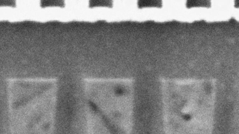
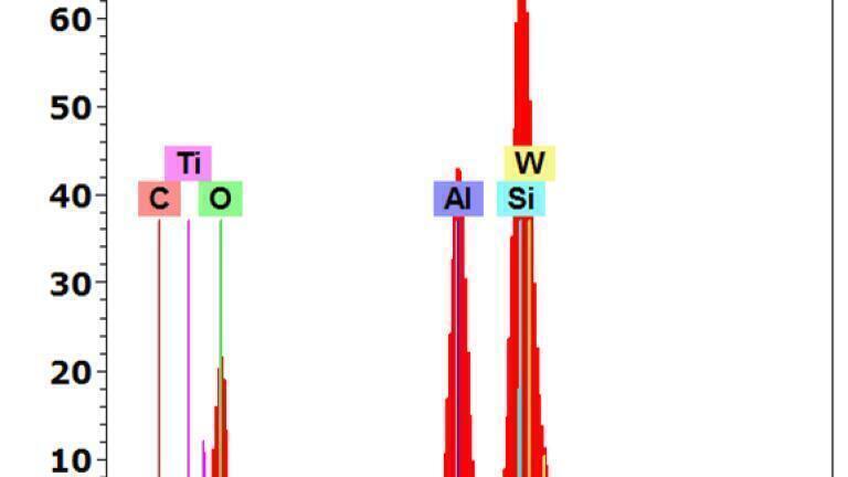
The analytical task in this case was to locate the element tungsten (W) within the structure depicted in a DDR-RAM located in the first figure of this article. If working with low acceleration energies consequently only low energy peaks of elements can be used. Many peak overlaps in this region make analysis difficult, especially in the application we have chosen (which is quite common to semiconductor analysis), as the sample spectrum the second figure illustrates.
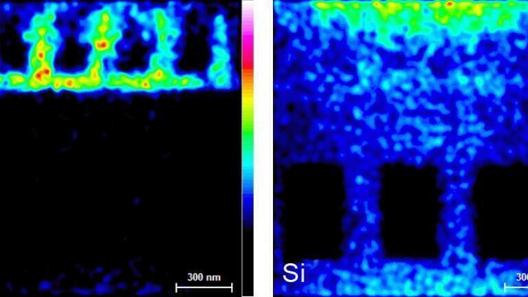
As can be seen, there is an almost 100% overlap between the tungsten M peak and the silicon (Si) K peak. This problem can be solved by a well-designed peak deconvolution procedure, which can accurately assign counts in a peak to either the one or the other element (works also with more than two elements). Mapping the whole sample and using peak deconvolution clearly separates W and Si, in the third figure shows.
It can therefore be concluded that tungsten is concentrated in the bright structure at the top of Fig. 1, while Si – as the substrate material – is present in most parts of the sample. This and similar information obtained can be an important aid in further improving semiconductor structures and to determine failures.
ILM 51.6 Sept 2026

 Product directory