-
 The skin was grafted onto mice
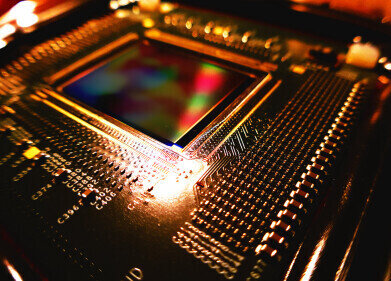
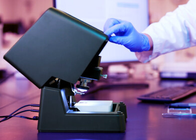
Microscopy & Microtechniques
Microscopy methods developed in fake skin experiment
Apr 22 2010
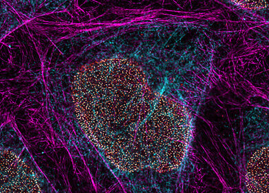
Laboratory scientists have developed new microscopy methods in order to examine fake tissue surgically added to mice.
Researchers at the University of Granada needed to introduce several inmunofluorescence techniques to enable experiments on the skin, Diligent Media Corporation reports.
The substitute membranes, created using tissular engineering, underwent quality control operations - including checks for cell proliferation, the expression of cytokeratin and the presence of differentiating morphological markers.
Following tests, the lab experts found satisfactory biocompatibility levels with the mice, with the skin showing no signs of infection or rejection.
According to the news provider, all of the animals used in the study exhibited granulation of the grafted tissue within six days, while nearly three weeks later cicatrisation - the forming of scars - had completed.
Earlier this month, the Engineer magazine reported that the latest microscopy procedures are being implemented in a Wellcome Laboratories study to develop more effective treatments for eye diseases.
Digital Edition
Lab Asia 31.2 April 2024
April 2024
In This Edition Chromatography Articles - Approaches to troubleshooting an SPE method for the analysis of oligonucleotides (pt i) - High-precision liquid flow processes demand full fluidic c...
View all digital editions
.jpg)


Events
May 05 2024 Seville, Spain
InformEx Zone at CPhl North America
May 07 2024 Pennsylvania, PA, USA
May 14 2024 Oklahoma City, OK, USA
May 15 2024 Birmingham, UK
May 21 2024 Lagos, Nigeria


