-
 Video microscopy was used to obtain results
Microscopy & Microtechniques
Microscopy used in cell division project
Apr 06 2010
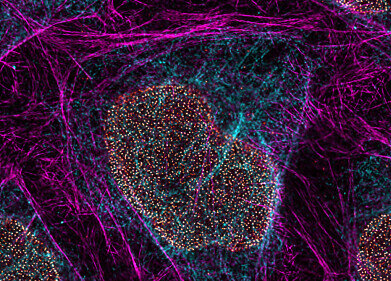
A six-year study that used microscopy to examine genetic cell division has now been published.
Findings from the MitoCheck project, coordinated by the Research Institute of Molecular Pathology (IMP) in Vienna, were outlined in the Science and Nature journals.
According to IMP, a facility founded in 1988, the process of cell division has "puzzled biologists for the past 150 years" and the work of researchers led by Jan-Michael Peters has come closer to solving the riddle.
Experts had to inactivate more than 22,000 genes in cultured cells in order to discover which ones were important for this process.
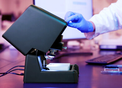
Video microscopy was then utilised to make movies of the samples to see whether powering down the individual organism units affected division.
"Our database is going to be an important source of information for many areas of biomedical research," Dr Peters suggested, adding that cooperation between international departments enabled the venture to be a success.
Findings from the MitoCheck project, coordinated by the Research Institute of Molecular Pathology (IMP) in Vienna, were outlined in the Science and Nature journals.
According to IMP, a facility founded in 1988, the process of cell division has "puzzled biologists for the past 150 years" and the work of researchers led by Jan-Michael Peters has come closer to solving the riddle.
Experts had to inactivate more than 22,000 genes in cultured cells in order to discover which ones were important for this process.
Video microscopy was then utilised to make movies of the samples to see whether powering down the individual organism units affected division.
"Our database is going to be an important source of information for many areas of biomedical research," Dr Peters suggested, adding that cooperation between international departments enabled the venture to be a success.
Digital Edition
Lab Asia 31.2 April 2024
April 2024
In This Edition Chromatography Articles - Approaches to troubleshooting an SPE method for the analysis of oligonucleotides (pt i) - High-precision liquid flow processes demand full fluidic c...
View all digital editions


.jpg)


Events
InformEx Zone at CPhl North America
May 07 2024 Pennsylvania, PA, USA
May 14 2024 Oklahoma City, OK, USA
May 15 2024 Birmingham, UK
May 21 2024 Lagos, Nigeria
May 22 2024 Basel, Switzerland


