Microscopy & microtechniques
Published over 9 years ago. See the latest and most current information on Microscopy & microtechniques.
Researchers are constantly striving for new ways to improve medical research. The Human Cell Atlas could do exactly that. If successful, the project is expected to contribute hugely to our understanding of how illnesses and diseases develop at cellular level. How? Read on for an introduction to what could be a massive research tool for scientists.
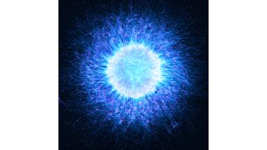
This isn’t your standard research assignment at a university, nor is it a few institutions working together. The Human Cell Atlas is an international project that will involve laboratories all over the world. There are an estimated 35 trillion cells in the human body, and researchers want to put together all the information they can gather about each and every one of them.
Whether the cells are diseased or perfectly normal, collectively the scientists are aiming to create an atlas of information about all of them and all their types. Why? It will give them a much deeper understanding of how diseases and mutations develop and spread.
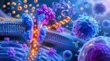
Cells are what all of our bodies are made up of. When they become abnormal, we get ill. Using what we currently know about cells, we can analyse, understand and consequently treat a huge number of diseases. But extending this to the vast number and types of cells in the human body essentially expands the foundation for much greater research.
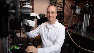
“It has become clear with new technology that there are lots and lots of cell types and cell states that we know nothing about. The Human Cell Atlas is about discovering what we’re missing,” explains Dr Sarah Teichmann, leader of the Sanger Institute’s gene expression genomics team.
According to Sanger’s website, the initiative was discussed at an international conference in London recently and will eventually be made freely available to scientists worldwide. The pilot studies contributing to the first phase of the project have already begun, looking at the immune system, nervous system, organ (epithelial) tissues and cancerous tumours. Looks at a new
There are a number of challenges the scientists face when gathering all of this information. One such challenge is capturing the dynamics of cells. While microscopic images allow scientists to analyse the cell’s properties, they don’t carry essential temporal information. ‘Flash-and-freeze Electron Microscopy – Adding Motion to Electron Micrographs’ looks at a new technique – ‘flash-and-freeze’ – which could provide scientists with the ‘missing piece’ in their research.
ILM Guide 2026/27

.jpg)