Microscopy & microtechniques
Published over 14 years ago. See the latest and most current information on Microscopy & microtechniques.
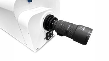
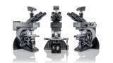
The University of Marburg is expanding its cooperation with Leica Microsystems: The Institute of Cytobiology is currently one of four institutes in the world to test a microscope with a resolution well below the diffraction limit (nanoscope). “With this new optical nanoscopy called GSDIM (ground state depletion microscopy followed by individual molecule return), resolutions down to 25 nanometers can be achieved. This makes it possible to image sub-cellular structures or protein complexes far beyond the resolving powers of a light microscope,” says cell biologist Professor Dr Ralf Jacob. The new technology, for which Leica Microsystems has been granted an exclusive licence, is being tested until September in the Imaging Core Facility of the special Cell Biology Research Department 593 (SFB 593) in Marburg. True-to-detail imaging of the spatial arrangement of proteins and other biomolecules in cells and observing molecular processes – GSDIM makes this possible for researchers due to resolutions beyond the diffraction limit. The
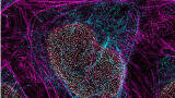
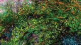
more insight science gains into these basic processes of life, the better it can find the causes of previously incurable diseases and develop suitable therapies. One of the strengths of GSDIM is that it uses conventional fluorescence markers to image proteins or other biomolecules within the cells with sharpness down to a few nanometers. This includes fluorophores that are routinely used in biomedicine.
With GSDIM, the fluorescent molecules in the specimen are almost completely switched off using laser light. However, individual molecules spontaneously return to the fluorescent state, while their neighbors remain non-illuminating. In this way, the signals of individual molecules can be acquired sequentially using a highly sensitive camera system and their spatial position in the specimen can be measured and stored. An extremely high-resolution image can then be created from the position of many thousands of molecules. This enables cell components that are situated very close to one another and cannot be resolved using conventional widefield fluorescence microscopy to be spatially separated and sharply reproduced in an image. With GSDIM technology, Leica Microsystems is extending its lead as an innovative provider of super-resolution light microscopes and nanoscopes. “With this new widefield microscope system we are extending our super-resolution portfolio and allow even more scientists to benefit from our innovative technology and advance their research,” comments Anja Schué from Leica Microsystems. The current test phase of the microscope is important in this context, as optimal testing can only be done by active scientists like the members of the SFB 593, which is sponsored by the German Research Society. “Our research naturally derives great benefit from being able to work with a microscope like this one,” added Professor Jacob
ILM 51.5 July 2026


.jpg)


-(1).jpg)