Chromatography
Published over 10 years ago. See the latest and most current information on Chromatography.
This article describes the use of Green Chemistry Principles when applied to the extraction and isolation of chlorophyll biodegradation products (Green Pigments) from pineapple peel waste. Green Pigments (GP) were isolated and identified from pineapple fruit peel extracts by using a renewable method based on a two phase extraction system (TPE) using ether/acetone at 40°C. The extract was worked up and purified via flash column chromatography (FCC) and the fractional components monitored by thin layer chromatography (TLC) at room temperature and identified utilising spectroscopic analysis tools such as UV-visible, FT-IR, 1H-NMR, 13C-NMR and GC-MS analysis. Two components of green pigments were identified, chlorophyll types a and b respectively with all solvents being recovered. The advantages of this method include environmental friendliness, low preparation cost, isolation, identification, and ease of detection.
The solid waste output in the world is growing day by day in volume and in toxicity. More and more everyday products contain chemicals put our environment in grave danger. This work was carried out using the most important principle of green chemistry, which is referred to as, ‘It is better to prevent waste material than to treat or clean up waste after it is formed’ [1].
The biodegradation of bioorganic solid waste involves millions of tons of fruit and plant waste every year. The biodegradation of fruit and plant waste comprises a series of biodegradation transitions that bring about changes in texture, metabolic changes and colour. Colour changes are as a result of chlorophyll biodegradation. Much research on Chlorophyll biodegradation has been performed and papers have been published in this field. Diapic N., et al extracted chlorophyll pigments from Parrotia persica, Hamamelis virginiana, Vitis vinirera var pinot noir, Prunus serrulata and Fothergilla gardenii autumnal leaves [2-7]. Ewa M. studied the molecular design and environmental determinants of plant color concentration [8]. Donghua l., et al studied the effect of some biomarker characters like Cr (VI) against Chlorophyll a, b ratio with their structure, composition and function [9]. The isolation of chlorophylls a and b from spinach leaves was performed employing both counter-current chromatography [10] and extraction processing [11]. Gautam S. S. et al and Lara R. A. Studied the amount and activity of bromelain present in the stem and fruit peel of the pineapple plant [12,13]. More recently Suman S. and Kaushik C. worked on identifying the proper dose of inorganic nitrogen fertiliser in relation to the growth stage and leaf colour chart [14].
Due to the importance of GP in human health the choice of Green pigment extraction from the waste of pineapple fruit peel was the main goal of this publication. Green pigment is a mixture of chemical components found in a plant that absorb light and utilise it in order to make energy in the plant. Moreover, the measurement of photosynthetic pigments can provide basic information on the physiological status of a plant [15]. This research was monitored and controlled using spectroscopic analysis including TLC, UV-visible, FT-IR, 1H-NMR, 13C-NMR and GC-MS analysis.
Materials and Methods [16]
1. Sample preparation:
Pineapple peels were collected from the market region in Hilla provenance. The leaves were washed and cut into small pieces, drying under vacuum, then crushed in a mortar and pestle for homogenisation. The samples were kept in a dry place at room temperature for further evaluation. (Figure 1a).
2. Extraction of pigment:
6 gm. of sample was prepared from pineapple peels (Figure 1a) and placed in a soxhlet tube thimble and extracted using three different solvents (methanol, acetone, and diethyl ether) respectively using a soxhlet apparatus (Figure 1b). The extraction time for each solvent was 45 minutes until the solvent became colourless (Figure 1c). The crude extract was directly transferred to a cooling centrifuge for about 10 minutes at 2000 rpm at 8°C. The extracted sample was then dried using anhydrous MgSO4 and the supernatant transferred to a lypholyser apparatus to remove all the excess solvent (after the drying process all the solvents were recovered with a yield of 85%). The best yields observed in case of acetone then ether and methanol were (255, 220, and 112 mg/5 gm.) extract respectively.
3. Analytical methods:
a. The crude extract of 90% acetone was tested by thin layer chromatography (TLC) using aluminium backed silica 60 (20×20 cm) TLC plates with F254 florescence indicator and developed using acetone/ether in ratios of 10:1, 10:3 and 10:5 as mobile phase. The best separation was observed using the (10:1) mobile phase. Three spots were clearly identified when the TLC plate was examined by an E-Graph Image Server 5 version 2.0.4 (ATTO Corporation). The retention factors (Rf) were 0.21, 0.28, and 0.52 respectively (Figure 2a).
b. The sample was then transferred to a 30 × 500 mm glass chromatography column packed with Silica gel (250 -400) mesh size (stationary phase). The mobile phase employed was acetone: ether in a (10:2) ratio (Figure 1c). The flow rate for elution was 3ml/ min. and 10 fractions were collected over 40 minutes and tested within TLC. Fractions 6 and 7 exhibited two different spots with Rf= 0.43 for A and 0.68 for B (Figure 2b). Removing the solvent under vacuum with cooling (lypholyser). An oily green pigment was observed for components A and B respectively
4. Equipment:
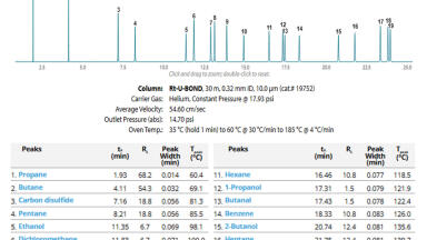
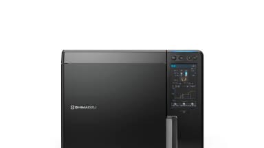
Various spectroscopic tools were used for identification in this application. The UV-Visible spectra were recorded on a Pye-Unicam 8700 spectrophotometer (200-900 nm), IR spectra were recorded on a Bruker Tensor 27 FT-IR spectrophotometer (4000-400 cm-1), using an ATR unit with Nujol mulls and liquid films between potassium bromide cells. E-Graph Image Sever 5 version with UV source 2.0.4 ATTO Corporation. Proton and Carbon Nuclear Magnetic Resonance (1H-NMR & 13C-NMR), Bruker, Ultra Shield Spectrometer 300 MHz, with tetra methyl silane (TMS) as standard, and CDCl3 + D2O as a solvent [18]. The Gas Chromatography – Mass Spectrometer (GC-MS), QP-2010 Ultra (Shimadzu Instrument, Japan) [19]. Column chromatography Glass Column 30×500 mm packed with silica gel 320-400 mesh, 1 gm./25 gm.) [17]. Freeze Dryer Christ, Alpha 1-4 LD, with Vacuum Pump RZ 6, up to 4-10-4 mbar.
Results and Discussion
The fundamental importance of this application was recycling impurities in the food waste from human nutrition to keep our environment safe and secure. The use of green chemistry principles were applied for the isolation and identification of one the most important biochemical pigments in living organisms. Historically, numerous methods were used for elucidation of the molecular structures of chlorophyll and its derivatives (Figure 3) [20]. More than four spectroscopic techniques were used in this research to prove and support the presence of GP in waste pineapple peels. UV-Vis spectra of the extract exhibit several absorptions bands (λmax) between 320-455 nm, indicating a mixture of several pigments, which were separated by column chromatography. Fourier transformer Infrared spectra (liquid disk and ATR mode) of the isolated compounds exhibit good evidence of the presence of oily GP type A and its derivatives (Figure 4). Two different types of carbonyl groups (ester & ketone) appear at two stretching frequency absorptions at n = 1737 and 1711 cm-1 respectively in rings I and IV. The conjugated system of the double bond C=C in the porphyrin isocyclic rings observed at stretching frequency n = 1463 cm-1. Large intensity of stretching absorption bands at n =2919 and 2862 cm-1 refer to the Ar-CH, CH2 and CH3 in the side chain groups and porphyrin ring. In addition C-O, C-N and CH bending which is exhibited at n = 1266 -1110 cm-1. Figure (4b) shows strong broad band stretching at
n = 3367 cm-1, OH carboxylic group, strong stretching absorption at n = 2020 -2850 cm-1, CH, aromatic and aliphatic groups, two types of stretching frequency absorption at n = 1736 and 1632 cm-1, carbonyl ester & keto respectively, and the rest of spectra for C=C conjugated, CH bending, C-N, and C-O appeared at 1465 -1381 and 1244-1070 cm-1 respectively.
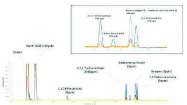
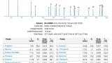
On the other hand 1H-NMR spectrum in CDCl3 (Figure 5a) showed the presence of Green Pigment type A as follows: three protons (Ha, Hb and Hc) of in the isocyclic porphyrin ring exhibited singlet at δ = 8.74, 8.49, and 8.25 ppm; three allylic protons bonded in C7 exhibited at δ = 8.07-8.09 ppm d, 1H, and 2H,dd at δ= 6.55-6.42 ppm respectively. Also one proton s, 1H of C19 at 6.16 ppm, two protons in C1 & C2 were exhibit at δ= 4.13 ppm d, 2H. Three types of four methyl groups bonding to C6, C11, C16, and C19 exhibited as follows; at δ= 3.69 ppm s, 2CH3, 6H refer to C6 and C16, s, CH3, in C11 at δ= 3.34 ppm, s, 3H at δ= 3.29 ppm refer to methoxy group, whereas the methyl group d, 3H at C2 exhibit at δ= 2.16 -2.11 ppm. The rest of signals belong to the ethyl in C12 pyrol ring II and phytyl side chain in C1 ring I. They were exhibited between δ= 2.56 to 1.50 ppm [21]. The other supporting evidences of isolation of GP type A was done by 13C-NMR in (Figure 5b). Two types of carbonyl C=O groups exhibited in down field as C=O, keto, IV ring at δ= 186.45 ppm and 2C=O, ester groups exhibited at δ = 174.53 ppm respectively. The other type of carbons such as C3, C18, C21, C5, C10, C8, C15, and C13 exhibited at δ= 160.33, 155.13, 154.02, 150.37, 148.11, 147.09, 146.23, and 140.04 ppm respectively. Also there are many signals with different intensity observed at δ= 139.58, 136.49, 130.85, 126.44 and 119.44 ppm which they belong to C12, C7, C6, C11 and C17 with carbon allylic group bonded at position C7. The rest of the signals clearly observed at δ= 65.10 to 22.91 refer to phytal side chain (R1, Figure: 3). From another side the elucidation of GP type B were done by GC-Mass analysis (Figure 6a &b) with the following condition, temperature injector programming 40 to 100ºC, column temperature 250-340ºC and the detector temperature was 350ºC. The chromatogram in Figure 6a exhibits a broad band width at 4.5-6.0 with peak height at 163. This provides good evidence and supports the presence of GP type b in its isolated form. Moreover the mass spectrum of the GP b exhibits molecular ion peak EI-MS+ at m/z = 636 ([M+Na]+), also additional signal exhibited at m/z= 617 ([M+H]+) fragment ion peak, and fragment ion at m/z= 592([M++H]-Mg) plus the rest of fragments at m/z= 504, 453, 335, 266 with base peak 200 respectively. Through these measurements the identification of isolated compounds matches 90% with literature publications [22].
Some applications of Green Pigment (GP)
Some of these publications suggest that GP is essential in photosynthesis and is instrumental in good human health.
1. GP helps control Hunger and promote losing weight [23].
2. It’s controls body odour by reduction odour, constipation and gas [24].
3. Encourages healing and helping in wounded therapy [25].
4. Removing toxic metals by binding with them to prevent absorption [26].
5. Protects DNA against fried foods [27], and potent antioxidant properties by protect cells from the oxidative damage by eliminating free radicals generation [28].
6. Effective against candida albicans growth [29]. Also able to reducing systematic redness and swelling [30].
7. Promotes healthy iron levels by new GP derivatives [31].
In addition to all these benefits, the waste of leafs (by-products) have a low commercial value, therefore can directly returned to the fields as soil improvements. Also the wastes are rich in the enzyme bromelain that could be isolated and purified in order to generate a product with a higher quality value.
References:
1. “The Principles of Green Chemistry”. United States Environmental Protection Agency. 2006-07-31.
2. Djapic, N. & Pavlovic, M. Chlorophyll Catabolite from Parrotia Persica Autumnal Leaves. Rev. Chim. (Bucuresti) (2008)., 59(8), 878.
3. Djapic, N, Pavlovic, M, Arsovski, S, & Vujic, G. Chlorophyll Biodegradation Product from Hamamelis virginiana Autumnal Leaves. Rev. Chim. (Bucuresti) (2009)., 60(4), 398.
4. Djapic, N, Djuric, A, & Pavlovic, A. Chlorophyll biodegradation in Vitis vinifera var pinot noir autumnal leaves. Research Journal of Agricultural Sciences (2009)., 41(2), 256.
5. Dapic N., facta universitatis, Physics Chemistry and Technology (2012) 10, 1, 21.
6. Ðapic, N. Behaviour of Fothergilla gardenii chlorophyll catabolite under acidic conditions. Kragujevac J. Sci. (2012),
34, 79.
7. Nina Djapic , Vasile Goldis University,Press (www.studiauniversitatis.ro) ,(2014), 24, 4, 314.
8. Ewa M?odziñska, Acta Biologica Cracoviensia Series Botanica , (2009), 51/1, 7.
9. Donghua l., Jinhua z., Keli y., Zhonggui z., and Wusheng j., Acta Biologica Cracoviensia Series Botanica, (2009) 1, 23.
10. Carole J and George B. Journal of Chromatography A, (2007), 1140 95; available online at www.sciencedirect.com.
11. Isabirye D.A., and Dikio E.D. * Bull. Chem. Soc. Ethiop. (2008), 22(2), 301. ISSN
12. Gautam S. S., Mishra S. K., Dash V., Amit K. Goyal and Rath G., Thai J. Pharm. Sci. (2010) 34, 67.
13. Lara R. A, Pereira B, Igor T. B., Edgar S.,Elias B. T., and Priscila G. M. , (2013) Braz. Arch. Biol. Technol. 56 6, 971.
14. Sarkar S. and Chakraborty K., Journal of Harmonized Research in Applied. Sciences, (2014), 2(4), 279.
15. Xueyun H., Ayumi T., and Ryouichi T., Bio.Med.Centeral, Plant Methods, Japan, (2013), 9,19.
16. Alaa J.M, Gafil H.S and Ehab K. A., Int. J. Chem. Sci.: (2015), 13(2), 983.
17. Lab of Research in Biochemistry department Collage of Medicine Babylon University Hilla Iraq.
18. Chemistry Department Collage of Science Al-AlByte University Irbid-Jordan.
19. Chemistry Department Collage of science Al-Mustansiriyah University Baghdad Iraq.
20. Nina Dapic , Kragujevac Journal of Science; (2012) 34, 79. UDC 581.19:582.638.28.
21. Cartelat A, Cerovic ZG, Goulas Y, Meyer S, Lelarge C., Prioul JL, Barbottin A, Jeuffroy MH, Gate P, Agati G.; Journal of Field Crops Research, (2005), 91.35.
22. Claudia Pellerito, Paolo D_Agati, Tiziana Fiore, Caterina Mansueto , Valentina Mansueto , Giancarlo Stocco, Laszlo
Nagy, Lorenzo Pellerito, Journal of Inorganic Biochemistry, (2005), 99, 1294.
23. Stenblom EL, Montelius C, Otbring K, Hakansson M, Nilsson S, Rehfeld JF, Erlanson-Albertsson C., Appetite. 2013
Sep; 68:118-23. doi: 10.1016/J.appet.2013.04.022. Epub 2013 Apr 28.
24. Young RW, Beregi JS Jr. Journal of American Geriatric Society, (1980);28(1):46.
25. Weir D, Farley KL Journal Wound Ostomy Continence Nurs. (2006); 33(5) :482.
26. Jubert C, Mata J, Bench G, Dashwood R, Pereira C, Tracewell W, Turteltaub K, Williams D, Bailey G., Cancer Prev
Res (Phila). (2009), 2(12):1015.
27. Shaughnessy DT, Gangarosa LM, Schliebe B, Umbach DM, Xu Z, MacIntosh B, Knize MG, Matthews PP, Swank AE,
Sandler RS, DeMarini DM, Taylor JA.,Journal Pone . (2011), 25,6.
28. (a).Zhang YL, Guan L, Zhou PH, Mao LJ, Zhao ZM, Li SQ, Xu XX, Cong CC, Zhu MX, Zhao JY., Zhonghua Nei Ke
Za Zhi. (2012); 51,6,:466.(b). El-Sayed WM, Hussin WA, Mahmoud AA, AlFredan MA. Biomed Res Int. (2013), 23,
10, 1155.
29. Lilian E. MAEKAWA, LAMPING R., MARCACCI S., Marcos Y. MAEKAWA, Maria R. Giazzi, Cristiane Y. Koga-Ito.
Revista Sul-brasiliera de Odontologia 01/2007.
30. Subramoniam A, Asha VV, Nair SA, Sasidharan SP, Sureshkumar PK, Rajendran KN, Karunagaran D, Ramalingam K., Inflammation. (2012); 35(3): 959.
31. Miret S, Tascioglu S, van der Burg M, Frenken L, Klaffke W. J. Agric Food Chem. (2010); 58(2): 1327.
ILM 51.5 July 2026

.jpg)
.jpg)


-(1).jpg)