Mass spectrometry & spectroscopy
A three-minute tissue analysis could provide surgeons with molecular evidence to distinguish glioma from non-tumour brain tissue and assess tumour characteristics, although its clinical value during surgery remains to be established, writes Alan Booth, Senior Editor, International Labmate.
Researchers at Purdue University and Mayo Clinic in the USA have developed a novel mass spectrometry method to analyse brain tissue in only three minutes. The approach could help surgical teams assess glioma margins and obtain molecular information within the time available for an operation, rather than needing to wait for subsequent laboratory results from biopsy.
The team evaluated the method in 86 human brain tissue samples and compared the analytical results with established clinical information. The findings have demonstrated its potential to distinguish glioma from non-tumour tissue and provide evidence about tumour subtype and mutation status. However, the measurements in this study took place away from the operating theatre. Further research will be required to establish whether the method can support surgical decisions and improve patient outcomes.
The research was published in September 2026 in the Proceedings of the National Academy of Sciences (PNAS). Dr Mahdiyeh Shahi, a former Purdue graduate student who is now a clinical chemistry postdoctoral fellow at the University of Minnesota Medical School, and Dr. R. Graham Cooks, the ‘Henry Bohn Hass’ Distinguished Professor of Chemistry at Purdue, led the work.
Cooks is based in the ‘James Tarpo Jr. and Margaret Tarpo’ department of chemistry at Purdue University in West Lafayette, Indiana. The collaboration also included Dr Alfredo Quiñones-Hinojosa, a professor of neurosurgery at Mayo Clinic in Jacksonville, Florida, his postdoctoral fellow Dr. Loizos Michaelides and other researchers at the Mayo Clinic.
Gliomas present a particular surgical challenge because tumour cells can infiltrate adjacent brain tissue rather than remain within a clearly defined boundary. Their molecular diversity also complicates diagnosis and treatment. Cells at the tumour margins may be difficult to identify with the technologies available during an operation, while residual disease can contribute to recurrence.
The surgical aim is therefore to remove as much tumour as safely possible while preserving healthy brain tissue and neurological function. More extensive removal is not automatically appropriate with the surgeon needing to consider the tumour’s location, the function of surrounding tissue and the patient’s overall clinical circumstances. A rapid molecular test could provide additional evidence to inform that judgement.
Conventional pathology and molecular investigations remain essential to diagnosis. However, detailed molecular results are not generally available within the few hours of an operation. The proposed method seeks to address this gap through rapid analysis of tissue already removed for assessment.
“The information we are getting is currently not available under standard of care procedures,” said Shahi.
“If our diagnostic method were to be approved, it would provide actionable molecular information during surgery. This information is currently not available on the time scale of the surgery.
“This information could directly influence treatment, including the extent of tumour resection, although the final surgical decision would be made by the surgeon based on the overall clinical context rather than the mass spectrometry results alone. It could also help guide the selection of targeted therapies,” she added.
Mass spectrometry measures the mass-to-charge ratio of ions, or electrically charged particles, to obtain information about a sample’s chemical composition. In biological tissue, it can detect molecules that reflect cellular metabolism and identify chemical differences between diseased and non-diseased regions.
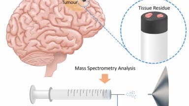
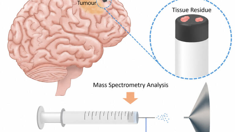
The researchers used syringe touch spray, an ambient ionisation technique designed to permit analysis with minimal sample preparation. Ambient ionisation produces ions under open environmental conditions that therefore do not require the sample to undergo an extensive preparatory procedure before analysis.
For this study, the researchers transferred small portions of brain tissue onto the plunger of a hypodermic syringe. A spray solution enabled the subsequent mass spectrometric measurement. The complete analytical process took around three minutes, a timescale that could make repeated tissue assessments feasible during surgery if the approach were to receive the appropriate clinical validation.
The method used tandem mass spectrometry, which involves successive stages of ion selection and analysis. This provides greater chemical specificity than a single mass measurement and helps reduce interference from compounds with similar masses or structures.
Rather than rely solely on an individual metabolite signal, the researchers calculated ratios between signals from pairs of endogenous compounds. Endogenous metabolites are substances produced or present naturally within the body. The relative signals can provide information about changes in tissue chemistry and offer an alternative to interpretation based on a single molecular measurement.
“The study used 86 human brain samples and the research showed that ratios of endogenous metabolites in brain tissue can distinguish glioma from non-tumour tissue with high confidence,” said Cooks.
“It can also classify tumour subtypes and mutation status. The simple ambient ionization mass spectrometry approach enables fast, clinically compatible analysis with potential to improve margin assessment and enhance patient outcomes,” he added.
The paper reported differences associated with tumour grade and the mutation status of isocitrate dehydrogenase, an enzyme involved in cellular metabolism. Mutations in this enzyme can cause the metabolite 2-hydroxyglutarate to accumulate. Such molecular differences help explain why metabolite measurements may provide diagnostic information beyond the tissue’s visible appearance.
Several metabolite ratios agreed completely with independent clinical classifications within the study dataset. That result does not establish perfect diagnostic accuracy in routine practice and so its performance will need to be assessed in further samples and under the conditions of an operation.
Cooks explained that the approach builds on ambient ionisation technology developed at Purdue. The first such method was described in 2004 and has since been commercialised and adopted for a range of analytical applications. Syringe touch spray extends this principle through a straightforward sample interface based on a conventional hypodermic syringe.
The tissue samples came from precision surgery guided by magnetic resonance imaging. Established diagnostic methods provided the reference information against which the mass spectrometry results were compared. This comparison allowed the researchers to assess whether the rapid chemical measurements reflected clinically recognised features of the samples.
The team now plans further work to assess the technique during surgery. According to Cooks, a study with more than 50 patients would be required as a next step and could provide the basis for a subsequent clinical trial. The current study comprised 86 tissue samples but this figure does not imply biopsy material from as many individual patients.
“In previous work, our measurements have been made in the hospital, during surgery,” said Cooks, with reference to research published in PNAS in 2024.
“In this case, we recorded the mass spectrometry remotely and compared with the available clinical information,” he said.
The distinction matters because analytical performance in collected tissue does not alone establish the practical value of a test in the operating theatre. Clinical assessment must determine how reliably the method performs within the surgical workflow and whether the information it provides can usefully inform decisions about tumour removal.
“This technology would not replace the current standard of care during brain tumour surgery,” said Cooks.
“Instead, it could provide surgeons with molecular information that is not currently available in the operating room, helping to guide surgical decisions during tumour removal, particularly at the tumour margins,” he concluded.
The specimens were supplied through the Mayo Clinic Florida Neurosurgery Biospecimens Repository of Intraoperative Patient Donations to Foster Collaborations Across the Globe and Enterprise (BRIDGE) Biobank, a central nervous system bioresource that provides patient-derived material for translational research. Such resources connect clinical specimens with research methods that may ultimately inform diagnosis and treatment.
Purdue’s chemistry department has an established record in analytical chemistry, chemical education and drug discovery, with two Nobel laureates in organic chemistry among its institutional distinctions. The wider university is a public, land-grant research institution with campuses that include West Lafayette and Indianapolis. Its background in analytical instrumentation provides the setting for this collaboration between chemistry researchers and clinical specialists.
The immediate contribution is a rapid method to extract molecular information from small brain tissue samples. Its potential clinical role is to supplement existing assessments at the point when surgeons must decide how far tumour removal can safely proceed. Prospective studies will be necessary before the technique’s effect on surgical practice or patient outcomes can be established.
The research team also included Dr. Diogo Moniz Garcia, a former Mayo Clinic postdoctoral fellow who is now a postgraduate neurosurgery resident at Washington University in St Louis, Missouri, and Aleeshba N. Basil of Mayo Clinic. Dr. Kenneth Lane Virgin of Indiana University School of Medicine contributed to the study and was previously affiliated with Purdue’s Aston Laboratories for Mass Spectrometry.
Shahi and Cooks are members of the Purdue Institute for Cancer Research and have worked within the university’s analytical chemistry research group.
Support came from the Purdue Institute for Cancer Research through National Institutes of Health grant P30CA023168 and the National Cancer Institute’s Innovative Molecular Analysis Technologies programme through grant R33CA240181-01A1. Further support came from the National Center for Advancing Translational Sciences and its New Chemistries for Undrugged Targets through A Specialized Platform for Innovative Research Exploration collaborative research programme, under grant UG3TR004139. Waters Corporation provided corporate support.
For further reading please visit: https://doi.org/10.1073/pnas.2616216123
LMUK 51.7 Nov 2026


 Product directory