Microscopy & microtechniques
Published over 6 years ago. See the latest and most current information on Microscopy & microtechniques.
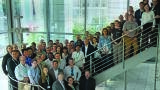
The international chemical imaging community gathered once again in Ulm, Germany, to share and discuss results, methods and technologies related to Raman microscopy. Almost one hundred attendees heard presentations arranged into distinct sessions under the headings: Nanotechnology and Low-dimensional Materials, Geosciences, Life Sciences, Applied Chemical Analysis, and Contributed Talks. Poster sessions provided a relaxed forum for browsing the many scientific results entered into this year’s Poster Award competition and a fascinating evening lecture looked at the entirety of progress in microscopy.
A grand total of seventeen presentations were delivered from the stage of the Ulm Stadthaus’s auditorium. Sebastian Schlücker (University of Duisburg-Essen, Germany) set the conference in motion with his introductory lecture on the theoretical background of Raman spectroscopy, including its classical and quantum mechanical descriptions, and its use in microscopy. An accompanying interactive quiz let the audience test their understanding of the physics behind the Raman effect. Olaf Hollricher (Managing Director-Research and Development, WITec GmbH) then detailed Raman imaging instrumentation, including spectrometers, detectors and software tools, in addition to describing considerations such as resolution and throughput. José Fernández (Instituto de Ceramica y Vidrio, CSIC, Madrid, Spain) then explored the combination of other methods with Raman imaging in his talk on correlative microscopy techniques.
Following the first poster session, it was time for the evening lecture. Charles Lyman, the Chief Editor of Microscopy Today, reviewed the course of innovation in microscopy while providing technological and historical context. He categorised advances as either revolutionary or evolutionary and reserved the term ‘breakthrough’ for those that enabled scientists to see something previously inaccessible and that led to the establishment of a new field of study. This comprehensive account of the development of microscopy closed out the first day of the Symposium.
The second day of the conference began with coffee and the fluent multidisciplinary exchange of ideas that the event has become known for. The first session of the day was dedicated to nanotechnology and low-dimensional materials. Yuan Huang (Chinese Academy of Sciences, Beijing, China) presented measurements of molybdenum disulphide and showed how Raman and photoluminescence can be used to monitor their strain-induced properties. Holger Schmalz (University of Bayreuth, Germany) showed how his work on electrospun multicomponent polymer fibres, particles and mesostructured systems benefits from Raman imaging. Nanostructured and composite membranes, Janus fibres and biohybrid microparticles were among the application examples he included in his talk. The session concluded with a presentation from Simon Thiele (Forschungszentrum Jülich and Helmholtz Institute Erlangen-Nürnberg for Renewable Energy, Erlangen, Germany) on serial section-based Raman tomography. The process of compiling 2D Raman scans into 3D images was memorably visualised with slices of marble cake.
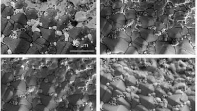
Geosciences were the focus of the next session, and two groups showed the audience the depth of the information that can be extracted from mineralogical samples. Maria Alexandrovna Sitnikova and Khulan Berkh (Federal Institute for Geosciences and Natural Resources, Hannover, Germany) offered a view into their research on distinguishing polymorphs using RISE (Raman Imaging and Scanning Electron) microscopy and included an example of 3D Raman imaging’s ability to reveal melted inclusions below a sample surface. Linda Prinsloo (University of the Witwatersrand, Johannesburg, South Africa) presented her work on prehistoric stone tools that provides insight into human technological development. She carefully observes the response of rocks to heat to understand the production processes used to create them. Trace substances such as fatty acids found on the tools also tell us about how our ancestors employed their technology.
The next session featured applications of Raman in the life sciences. Katja Schenke-Layland (University of Tübingen, Germany) began with her investigations of engineered tissues for personalised medicine and monitoring of phenotype switching, including examples of smooth muscle cells for cardiovascular tissue and pseudo-islets expressing insulin trapped on a chip. Peter Vikesland (Virginia Tech, Blacksburg, USA) detailed the method of surface-enhanced Raman scattering (SERS) and described how surface plasmon enhanced elastic scattering signals can be used as internal standards to compensate for the signal heterogeneity of SERS substrates.
Applied chemical analysis was the common thread of the next session and Lars Meyer (BASF SE, Ludwigshafen, Germany) started it off with a medley of analytical tasks that he addresses in his industrial laboratory using Raman imaging. For example, he showed how gypsum crystallisation can be monitored and its rate of formation regulated through polymer additives. Erik Emmons (US Army Research Laboratory, Aberdeen, USA) employs Raman microscopy for the analysis of samples relating to chemical, biological and explosives defence. His talk featured results of explosives detection from fingerprints and showed the ability of Raman microscopy to distinguish between viable and deactivated biological spores.
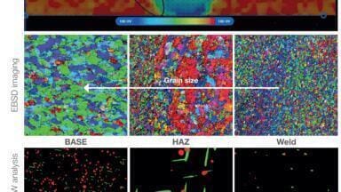
The contributed talks that concluded the second day at the Ulm Stadthaus were dispatches right from the forefront of Raman technology. Patrick Altmann (attocube systems AG, Haar, Germany) presented several examples of cryogenic Raman spectroscopy and imaging in high magnetic fields for research on low-dimensional materials and showed how the Raman spectra of a single graphene flake can be affected by changes in magnetic field strength. Bastian Barton (Fraunhofer Institute for Structural Durability and System Reliability LBF, Darmstadt, Germany) explained how he uses 3D Raman imaging for analysing multicomponent polymers and the ways in which additives can serve as flame retardants and structural reinforcements. An example he provided was a 3D representation of the core-shell structure of lubricant-containing microcapsules embedded in a polymer matrix. Emil Bjerglund (Danish Technological Institute, Aarhus, Denmark) gave a talk that concerned the detection of document fraud. He showed how forged receipts can be revealed by using Raman spectroscopy to chemically analyse the ink, as its spectra change slightly over time. The non-destructive and label-free nature of the technique is essential for this application. Armin Zankel (Graz University of Technology, Austria) wrapped up the session with a look at the combination of Raman imaging, electron microscopy (RISE) and energy-dispersive X-ray spectroscopy. His example measurements from materials science included vivid depictions of polymers and several mineralogical samples.
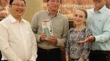
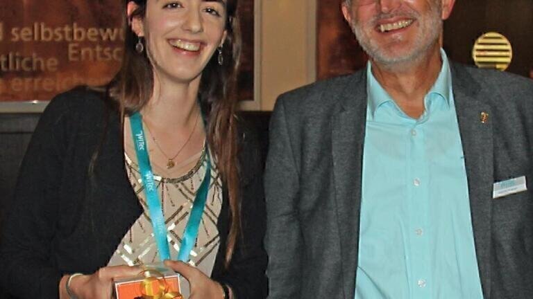
As the day turned to evening the attendees reconvened in the historic City Hall of Ulm for the conference dinner and the announcement of the winner of the Poster Award. Locally brewed refreshments and traditional Swabian culinary specialties accompanied discussions of the research presented. The competition between twenty-five scientific posters was intense and many favourites emerged among the guests. The jury had the difficult task of choosing only one and ultimately selected Birgit Bräuer of the University of Vienna as the winner of the 2019 WITec Poster Award. Her submission, Surface Characterization of Escherichia coli-imprinted Polymers using Confocal Raman Microscopy, is a striking demonstration of the utility and versatility of the technique in establishing the presence or absence of bacteria in a sample.
The third day of the Symposium brought the attendees from the city centre up to WITec headquarters for demonstrations of the very latest in confocal Raman imaging and correlative microscopy instrumentation. For many, it was their first opportunity to see technologies such as the ParticleScout microparticle analysis tool in action.
The 16th Confocal Raman Imaging Symposium was once again a resounding success, with the variety of application fields represented and the balance between multidisciplinary appeal and scientific depth maintaining the exceptional standard established by previous years.
The 17th Confocal Raman Imaging Symposium will take place from 28th - 30th September 2020.
More information online: www.witec.de
ILM 51.5 July 2026


-(1).jpg)
.jpg)
.jpg)

.jpg)