Research news
A flexible micro-endoscope shows promise for minimally invasive diagnostics deep within the body
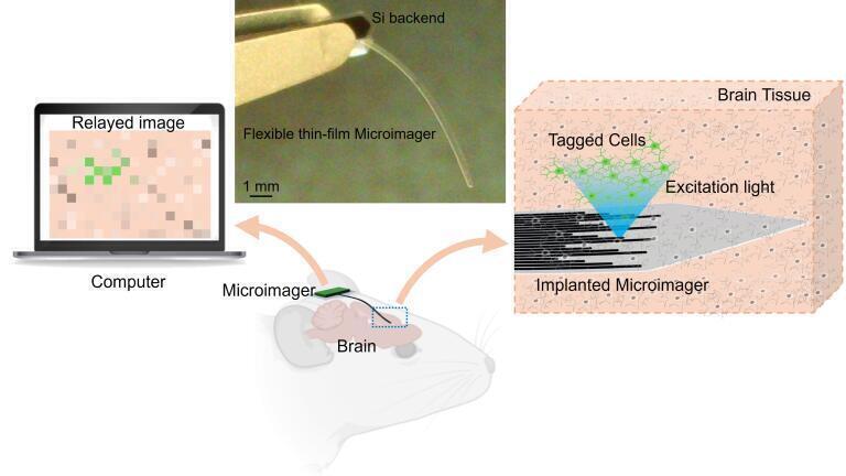
Researchers at Carnegie Mellon University have developed an exceptionally thin and flexible imaging device, potentially enabling the minimally invasive visualisation of internal tissues, including brain structures. The microimager, far smaller than a human eyelash, represents a major advance in endoscopic technology and may one day support early and accurate disease detection.
The device measures just 7 microns thick — around one-tenth the diameter of an eyelash — and 10 millimetres long. Its size and flexibility make it ideal for reaching delicate and deep regions of the body without causing significant damage to surrounding tissue.
“Unlike existing endoscopes, which rely on large camera systems or bulky fibre-optic bundles, our microimager is highly compact and well-suited for minimally invasive procedures,” said Professor Maysam Chamanzar, who led the research team. “This makes it feasible to image parts of the body that are currently difficult or impossible to access without significant tissue disruption.”
In laboratory demonstrations, the team successfully used the imager to visualise brain structures and neural activity in live mouse models. The imaging platform is based on a flexible photonic system constructed from Parylene—a biocompatible and transparent polymer already used in implantable medical devices. The researchers harnessed Parylene’s ability to guide light bidirectionally to design waveguides capable of both delivering illumination and capturing scattered light from tissue.
Each waveguide in the device features micromirrors at either end. Some waveguides emit light to illuminate biological tissue, while others collect the backscattered light, which is then transmitted to an image sensor. Each waveguide essentially functions as a pixel in the final image.
“We fabricated the device using microscale processes akin to those used in the production of microelectronics and MEMS,” explained M. Hassan Malekoshoaraie, a doctoral student who led the design and experimental validation of the endoscope. “This allows us to tailor the waveguide arrangement to suit different tissues or imaging requirements.”
The researchers validated the system by imaging fluorescent beads in a scattering medium to demonstrate 3D localisation. They then captured fluorescence images from mouse brain tissue expressing green fluorescent protein and further demonstrated the device’s capacity for functional imaging using calcium indicators—markers of neural activity.
“To confirm the reliability of our optical imaging, we compared our results to ground truth electrophysiological recordings,” said neuroscientist Dr Vishal Jain. “The close correspondence between the datasets gives us confidence in the accuracy of our imaging technique.”
Professor Chamanzar added that the team’s long-term objective is to map neural activity alongside gene expression in specific cell types—an ambitious goal in systems neuroscience.
Future development will focus on integrating image sensors, filters and light sources into the back end of the device to create a fully self-contained platform suitable for in vivo use. Potential clinical applications include detecting residual cancer cells post-surgery and monitoring disease progression or recurrence.
“This microimager opens the door to a host of new possibilities in diagnostics and real-time surgical guidance,” Chamanzar said. “With further refinement, it could become an essential tool for minimally invasive medicine.”
For further reading please visit: 10.1364/BOE.558778
ILM Guide 2026/27


.jpg)
.jpg)


2.jpg)
