Microscopy & Microtechniques
Extensive Digital Imaging Features Enable Better Microscopy
Jan 28 2009
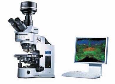
Olympus has launched the new and improved cell family range of mutually compatible imaging systems, combining excellent performance with user-friendly operation. These three software imaging programmes bring increasing numbers of capabilities to imaging systems so that each task, project or experiment can be completed using the most suitable set of tools.
cell^D is a comprehensive and reliable imaging and documentation system for life science microscopy. In conjunction with a digital camera and a microscope cell^D becomes the perfect system for microscope image acquisition, archiving and documentation. Images of unlimited depth of focus can be generated using the extended focus function either manually or via a motorised focus drive.
The cell^D software controls all necessary hardware, and contains a range of enhanced image acquisition tools, standard image processing and measurement functions. This is all
complemented by a structured database and a report generation tool.
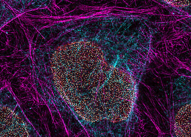
Olympus cell^F is designed with all the features of cell^D and includes the additional benefits of speciality tools for fluorescence imaging. It is therefore designed to handle automated
processes and multi-dimensional fluorescence image manipulation. cell^F is flexible and easily adapted to suit each researcher’s fluorescence acquisition, processing, visualisation and
documentation needs. Furthermore, it includes advanced spectral unmixing algorithms to separate fluorescence image information from multiple GFP variants enabling, for instance,
GFP and YFP to be used in the same fluorescence study. What is more, predefined position masks can be used to generate images of the experiment results in multiwell-plates.
Building on cell^F, cell^P is the software solution to meet the more demanding procedures within the field of life science imaging. Complex multiple fluorescence image-analysis tasks such as ratio imaging, delta F/F measurements or kinetic evaluation are completed with ease and can be carried out automatically. It also enables time-lapse imaging, out-of-focus haze removal and advanced image visualisation. Olympus cell^P can be integrated with other software platforms via the Imaging C scripting language, which enables more efficient imaging laboratory workflows.
If needs change, they can all be upgradeable to more advanced packages in the Olympus cell imaging family.
Digital Edition
Lab Asia 31.2 April 2024
April 2024
In This Edition Chromatography Articles - Approaches to troubleshooting an SPE method for the analysis of oligonucleotides (pt i) - High-precision liquid flow processes demand full fluidic c...
View all digital editions
.jpg)


Events
May 05 2024 Seville, Spain
InformEx Zone at CPhl North America
May 07 2024 Pennsylvania, PA, USA
May 14 2024 Oklahoma City, OK, USA
May 15 2024 Birmingham, UK
May 21 2024 Lagos, Nigeria


