Microscopy & microtechniques
Published over 10 years ago. See the latest and most current information on Microscopy & microtechniques.
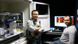
The Weatherall Institute of Molecular Medicine (WIMM) at Oxford University comprises various groups involved in molecular medicine research forming a ‘Research Hotel’. They all come from different medical departments and also different topics, but their joint efforts within one place and the possibility to use state-of-the-art facilities shall optimise research efforts and bolster novel results.
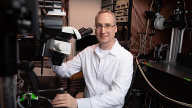
Dr Marco Fritzsche works in the Nano-Immunology group of Professor Christian Eggeling in the MRC Human Immunology Unit (HIU) at the WIMM. Their specialty areas include cell mechanics and the use of FRAP.
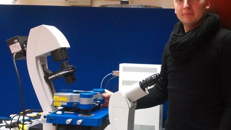
In recent years, biomedical researchers have increasingly recognised the importance of forces within biological systems, ranging from the spreading of cancerous cells to the triggering of the immune system resulting in the atomic force microscope (AFM) being adopted by many research groups on a daily basis. However, to date, no system had been available within the Oxford microscope facilities serving biological research for directly measuring or enforcing forces on cells. The way cells interact with each other is often ruled by physical changes due to motion, so-called mechanical force. Such forces are present during molecular rearrangements and are observed most obviously when cells form close contacts, avoid each other or reshape to change morphology. Forces play an important role in many biomedical processes. The new JPK AFM setup will eventually be integrated into the image facility at the WIMM, offering the possibility for less-experienced users to address questions about force involvement in their biomedical research.
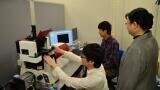
Dr Fritzsche and the Nano-Immunology group use a toolbox of different nanoscopy and microscopy techniques. This involves custom-built and commercially available microscopes such as Stimulated Emission Depletion (STED) microscopy, STORM, as well as confocal scanning, spinning disk microscopy, widefield technologies and TIRF. Moreover, they have implemented quantitative microscopy and spectroscopy methods such as FRAP, point FCS, scanning FCS, FLIM, and FRET.
Dr Fritzsche sees several benefits in choosing JPK´s system: “What is particularly important for our experiments is the ability of the JPK AFM NanoWizard® to move the sample instead of the head and the ability to pull and push with a dynamic range of 15 µm. This way, we can quantify the mechanical properties of the cells but also functionalise cantilevers with antibodies and receptor ligands and study the forces exerted by the immune cells in response to these ligands.”
ILM Guide 2026/27

.jpg)