Microscopy & microtechniques
Published over 11 years ago. See the latest and most current information on Microscopy & microtechniques.
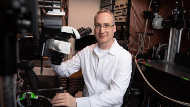
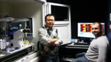
Dr Yves Dufrêne is a senior research fellow who is leading the Nanobio Team at UCL, Université catholique de Louvain. His research is at the cross-roads of nanotechnology and microbiology where the goal is to gain insight into the nanometer-scale surface architecture and molecular interactions of microbes. Two main topics are: understanding the spatial organisation of the cell wall and deciphering the molecular mechanisms of cell adhesion.
Looking more deeply at his work, Dr Dufrêne said: “the completion of our multidisciplinary research requires a novel, state-of-the-art AFM for the nanoscale analysis of live cells hence our choice of the NanoWizard® from JPK Instruments. This is equipped with a series of advanced capabilities that are essential for the project. First, the combination of AFM with a high-quality inverted optical microscope will allows us to optically guide AFM analyses (selection of a cell of interest, followed by targeted AFM measurements), therefore improving dramatically the throughput and quality of single-cell analyses. In addition, this AFM-optical microscopy platform will make it possible to directly correlate AFM topographic images with fluorescence images and to prepare living cell probes for single-cell force spectroscopy (SCFS) measurements. Secondly, the new high-speed module will complement traditional topographic imaging of bacterial cell surfaces by providing a more dynamic view of the cell surface (cell surface organisation, relaxation and turnover; cell wall remodelling in response to drugs). We believe this project is a unique opportunity to push the limits of high-speed AFM in microbiology. Third, an advanced single-molecule force spectroscopy (SMFS) package will enable us to image and manipulate, at high resolution, single cell surface constituents. This package will include the force-clamp mode, which will provide novel insights into the adhesive and mechanical properties of the adhesins. Fourth, the newly developed multiparametric imaging mode (also named quantitative imaging, QI™) will allow us to dramatically increase the spatiotemporal resolution of SMFS-based imaging, and to correlate the localisation of single cell surface constituents with local changes in adhesion and elasticity. Fifth, the unique cell adhesion force module (CellHesion™ module) will guarantee reliable, state-of-the art single-cell force spectroscopy (SCFS) measurements, and will permit, for the first time, long-range z-spectroscopy (up to 100 µm), thereby enabling to unravel molecular interactions that were not accessible before.”
ILM Guide 2026/27