Microscopy & microtechniques
Published over 10 years ago. See the latest and most current information on Microscopy & microtechniques.
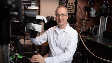
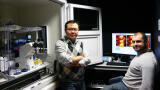
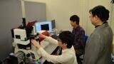
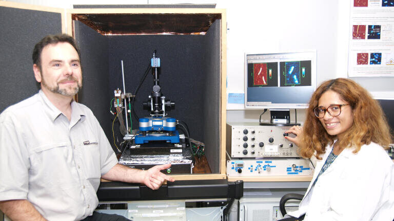
Drs Christophe Demaille and Agnès Anne from CNRS work in the Laboratoire d'Electrochimie Moléculaire at the Université Paris Diderot as Group Leaders of a research team aiming to probe electron transport and communication in nanometric biostructures. Their studies are performed at the single nano-object scale using combined atomic force (AFM)-Electrochemical (SECM) microscopy. In this combined microscopy technique, the tip acts as both force sensor and microelectrode. The Group produces their own hybrid AFM-SECM tips from gold microwires.
Dr Demaille says that their approach is unique in that they are using a particular AFM-SECM configuration, they labelled Mt (Molecule touching) AFM-SECM, where the tip comes in direct physical contact with the nano-object to be electrochemically interrogated. Their aim is to localise individual redox active biomacromolecules and probing their activity. This involves redox labelling of the structures of interest and requires both a nanometer range spatial resolution and the ability to measure very low electrochemical currents.
Recently in a collaborative work with the team of Thierry Michon from INRA, this approach has been successfully used to characterise nanometer sized biological objects such as viruses. The use of redox-immunomarking (redox labelled antibody targeting specific viral proteins) allows selective detection of the sought viral proteins since only the immunomarked proteins are seen in the tip current image. The virus topography is obtained simultaneously so that the location of the viral proteins can be resolved with respect to the virus external architecture.
Talking about their choice of system, Dr Demaille said: “We believe our NanoWizard-based AFM-SECM microscope is a promising tool for viral nanotechnology since it uniquely allows the functional characterisation of modified viruses. Historically, we started working on early model of commercial AFM which we modified to enable electrochemical measurements. Then we purchased the JPK NanoWizard® II system to specifically work on the ‘virus’ project with a budget from an ANR grant application. The JPK AFM is much easier to handle and to modify, especially with respect to the open access (to the cell) which is not possible with other tools from the competition. We have basically kept the prism and added a wire connected to a home-designed bipotentiostat. The new setup has been implemented in the software. What is also unique with the JPK setup is the presence of a real reference electrode which is the key requirement for this type of measurement.”
ILM Guide 2026/27