Microscopy & microtechniques
Published over 11 years ago. See the latest and most current information on Microscopy & microtechniques.
Researchers at Maastricht University Medical Center+ (Maastricht UMC+) and the MAASTRO Clinic have developed a new medical imaging analysis method to predict the risk of dying of cancer in patients. For the first time, they have been able to make a visual fingerprint of a tumour that provides detailed information. The new technique is known as radiomics and it makes use of standard imaging technology present in every hospital. Based on the specific information acquired with radiomics, doctors can select and apply the most effective treatment for each patient.
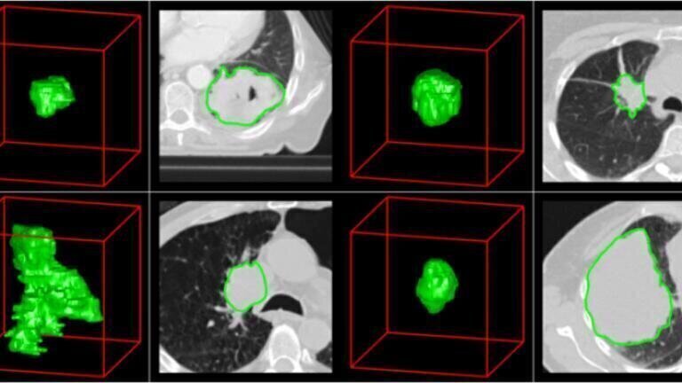
Tumours can differ substantially in terms of their properties, for example their shape, size and/or composition. Medical imaging technologies, such as a CT scan, offer a simple way of making some of these properties visible. In itself, however, the image says nothing about how the tumour will develop. Now, researchers have shown that the new radiomics technique can predict tumour progress. The researchers are participating in a major international study headed jointly by Maastricht UMC+ and Harvard Medical School in Boston (US).
The researchers quantified 440 properties taken from a total of 1019 CT scans of lung cancer and head and neck cancer patients. This involved using sophisticated and intelligent software that transforms the tumour properties shown in a scan into clinically meaningful numbers, giving rise to a kind of blueprint for that specific tumour. "That data allows us to unravel the digital fingerprint of each separate tumour, in a manner of speaking,” says Dr Philippe Lambin, professor of radiotherapy at Maastricht UMC+ and project leader. “We discovered that a subset of the lung tumours shared four characteristics related to intra-tumour heterogeneity and these cancers grew and proliferated faster than others, leading to worse patient outcomes. What really surprised us is that the exact same four characteristics - the same signature, if you like - showed up in aggressive, head and neck cancers too, again predicting a poorer survival after radiation therapy. That’s exceptionally valuable information that we can use to predict the progress of the cancer, among other things. We can now identify patients who will benefit from stronger medication, for example, while keeping treatment simple, pain-free and cost-effective."
The researchers are currently engaged in software development that will make the technique readily available to clinics worldwide at low cost. "The software doesn’t require any additional knowledge of medical imaging techniques, so it’s an ideal complement to existing hospital technology,” says clinical physicist André Dekker of the MAASTRO Clinic. The pharmaceutical industry has already taken a keen interest in radiomics.
Dr Hugo Aerts of Maastricht UMC+, who is currently conducting research at Harvard University, explains why: "When we can get detailed information about a tumour before commencing treatment, we can tailor that treatment to the individual patients. This 'personalized medicine' approach can result in much more effective treatment. That’s why radiomics could have a major influence on the development of personalized medicine in the field of protontherapy as well as the pharmaceutical industry."
Radboud University Medical Centre in Nijmegen, VU Medical Centre in Amsterdam and two institutes in the US, Moffit Cancer Center in Florida and Harvard Medical School in Boston, collaborated on the study. The research was funded by the Netherlands Cancer Institute, the European Union and the National Institutes of Health in the US.
The researchers have published the promising results of their study in Nature Communications.
ILM Guide 2026/27