
Microscopy & microtechniques
Published over 10 years ago. See the latest and most current information on Microscopy & microtechniques.
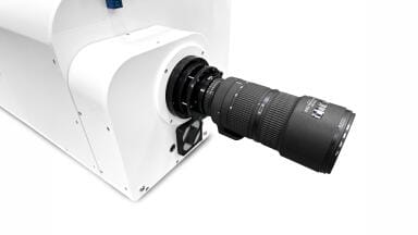
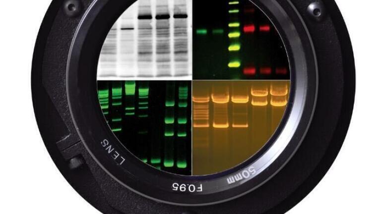
The imager features a choice of deeply cooled (-57°C) cameras for low noise, image capture in long exposure applications. Choose from a 2.1 MP camera with an f/1.2 zoom lens, or an 8.1 MP camera with an, f/0.95 lens, for fast capture. Lenses are offered with the choice of manual or motorized control. The darkroom additionally includes a five position, motorized filter wheel and a height adjustable tray, for imaging blots closer to the lens.
Add a BioLite MultiSpectral Light Source for RGB, NIR and multiplex imaging applications. The BioLite supplies overhead excitation of fluorescent multiplexed Western blots, DIGE 2D gels, PAGE gels and more. Filters are available for specific wavelength requirements.
Applications include fluorescent Western blotting, immunoblots, chemiluminescent blots (such as Western Blots, Northern Blots, Southern Blots), protein blots, multiplex imaging of multiple proteins in a single blot, wide range of stained gels, gel imaging and gel documentation, 1D quantitative analysis, Western blot area density analysis.
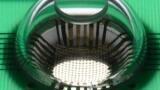
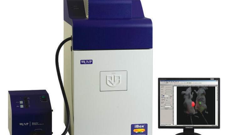
The imager features UVP’s new, deeply-cooled BioCam 900 camera (-90°C) with superior specifications for quick and sensitive capture. The darkroom contains a software controlled, height adjustable platform for imaging closer to the lens. System control, image capture and quantitative analysis are performed using UVP’s full package software VisionWorks LS. This sophisticated package provides comprehensive tools for image capture and analysis of gels, plates and membranes as well as colony counting.
An anesthesia delivery system is available for real-time imaging of small animals. A warming plate is available to provide a platform for maintaining a regulated temperature of small animals to prevent hypothermia. The stable temperature ensures consistent day-to-day results.
Additional UVP products include ultraviolet lamps and UV viewing cabinets, HEPA/UV PCR hoods, precision heated hybridization ovens and UV crosslinkers.
ILM Guide 2026/27

.jpg)