Laboratory products
By turning glass slides into shareable digital images, digital pathology could give patients in rural and underserved communities the same access to subspeciality expertise as those at major academic medical centres.
Pathology sits at the heart of modern medicine. Nearly every cancer diagnosis, treatment decision and patient journey through serious illness passes through the pathologist’s hands. Yet for decades, the quality of that diagnosis has often depended on geography – where a patient lives has determined who examines their tissue samples and how quickly they will receive the answers that they need. A rural community hospital might wait days for results that a patient nearer to a major academic medical centre can receive in only hours. They might have their case reviewed by a generalist when a subspecialist’s expertise could be transformative.
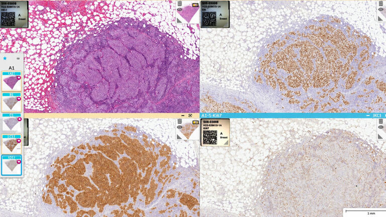
Digital pathology is fundamentally changing this equation by converting glass slides into high-resolution digital images that can be viewed, shared and analysed anywhere in the world. This technology promises to decouple diagnostic excellence from a clinic’s physical location. The implications extend far beyond convenience – this represents a potential restructuring of how healthcare systems deliver pathology services – with profound impacts for patients in underserved communities.
The pathology community has reached consensus on the essential point that digital transformation is inevitable, with the question being no longer whether laboratories will adopt digital pathology but how quickly and how completely they will move to make the transition.
Yet widespread routine adoption remains in its early stages. Today, the majority of pathology-based diagnostics worldwide are performed in the traditional manner with a pathologist sitting at a microscope, examining glass slide samples one-by-one. This persistence of analogue methodology exists despite widespread knowledge of digital pathology’s potential benefits.
The barriers to faster adoption are multifactorial. In the USA, the absence of specific reimbursement for digital pathology makes calculating a return on investment difficult to achieve within current healthcare settings. Benefits tend to be viewed as subjective and measured in savings and cost reduction rather than revenue generation. And so, administrators who are accustomed to assessing clear financial justifications find themselves evaluating a technology whose advantages are real but harder to quantify in traditional terms.
Beyond the economic questions lie the challenges of change management. Digitising pathology represents a full digital transformation project, requiring significant effort from multiple departments that are typically already understaffed. Both pathologists and laboratory technicians often find themselves stretched thin, being time poor in order to invest in the improvements that would ultimately make their labs more efficient.
Generational and psychological dimensions should also be considered. Pathologists who have trained for years or decades at the laboratory microscope may feel reluctant to abandon familiar tools, their muscle memory and tried-and-tested workflows. The transition requires not just the installation of new equipment but also acquiring new habits coupled with new modes of work.
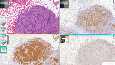
Digital pathology workflows begin with the process of scanning traditional glass slides in order to convert them into high-resolution digital images. Modern scanners can process slides rapidly, and create images detailed enough to support the same diagnostic decisions pathologists have always made but with amplified capabilities that glass slides cannot provide. Multiple systematic reviews, meta-analyses, and large-scale validation studies demonstrate that digital pathology achieves diagnostic concordance with conventional microscopy of approximately 97 to 99 per cent, supporting its use for routine primary diagnosis when appropriately validated. [1,2 3,4]
Once digitised, these images can be stored, transmitted, and viewed on computer screens anywhere that has adequate levels of internet connectivity. A pathologist sitting in a clinic in one city could also examine tissue from a patient whose location is hundreds or thousands of miles away just as easily as though they were reviewing a case from the same building. Multiple experts can view the same case simultaneously, collaborating in real time despite potentially vast geographic separation.
The technology has matured significantly in recent years with regulatory clearances having expanded. The USA’s Food and Drug Administration (FDA) has now cleared several systems to be available for primary diagnosis within the USA. [5,6] Integration with laboratory information systems allows digital pathology to fit within existing clinical workflows rather than requiring parallel processes.
Perhaps most significantly, the emergence of comprehensive end-to-end solutions means healthcare systems can now implement digital pathology without the need to match up components from multiple vendors. This new level of integration reduces complexity at implementation and should support smoother adoption in the lab itself.
The most compelling case today for digital pathology centres not just on efficiency or cost savings but on the opportunity to fundamentally improve patient care by eliminating geographic barriers to allow faster access to global expertise.
The current reality in many healthcare settings is that when a pathologist encounters a difficult case and decides they require a second opinion, practical constraints often determine who can provide that consultation. Traditionally, a pathologist with a challenging case might reach out to another pathologist who is on duty at their clinic for rapid access to a second opinion.
But this approach – choosing consultants based on convenience rather than expertise – represents a compromise that patients may never see but one that could impact their care. A generalist pathologist happens to be available so the generalist provides the second opinion, even when a subspecialist’s input could have proved to be more valuable.
And when a second opinion requires review by a specialist who is not onsite, fragile glass slides must be carefully packaged, paired with the relevant documentation, and couriered to that specialist, whether across town, across the country or elsewhere. The slides must then be received, unpacked, analysed, documented, repacked and returned to the originating institution. In addition to the administrative burden and cost, this process can delay diagnosis and treatment initiation by days – or even weeks – while introducing unnecessary risk of slide damage, loss, or other transit-related complications. Just think about the risks that simply the winter season can introduce.
Digital pathology eliminates these compromises given that when a case exists as a digital file rather than a physical glass slide geography becomes a meaningless factor in who could provide the first or second level of consultation.
A pathologist can potentially seek input from specialists anywhere even if they are hours and cities away.
This capability transforms treatment available at a smaller and more remote facilities for patients in less-served communities. They will no longer have to face the choice between travelling away from home – potentially when seriously unwell – and accessing top level expertise, especially to be able to receive subspecialised diagnostic input.
Large health systems and laboratories are beginning to implement digital pathology at scale, purchasing multiple scanners to transform their diagnostic operations. This trend represents an emerging model that could reshape pathology services more broadly.
The strategic logic involves centralising laboratory operations while simultaneously expanding diagnostic capabilities across the network. Physical specimens and scanning infrastructure can be consolidated for efficiency, while the resulting digital images flow freely to pathologists who are networked into the system and potentially beyond it.
This model offers scalability advantages over traditional approaches. Academic teaching hospitals have long served as centres of excellence in pathology but their usual model – concentrated expertise at a single location – naturally limits the numbers of patients they serve in their catchment orbit and requires patients to come to them. A distributed digital model can extend subspeciality expertise across entire health systems, reaching patients wherever they receive care.
Healthcare systems, including major academic institutions and large integrated delivery networks, have begun investing more heavily in digital pathology infrastructure than ever before, suggesting growing confidence in this approach.
Beyond patient care, digital pathology addresses the growing challenge of workforce sustainability in pathology. The field has faced shortages and laboratories must compete to attract and retain qualified pathologists. [7,8]
Digital pathology is emerging as a differentiator in this competition. Many pathologists who have adopted digital workflows increasingly report high satisfaction with the transition and would prefer not to find work in an organisation that is not digital. [9] For these practitioners, digital pathology represents not just a different way of working but a better one – more flexible, more connected, more aligned with how they want to conduct their professional practice.
The technology also shows promise for attracting new talent into the field. Recent discussions at professional meetings highlighted how exposure to digital pathology and artificial intelligence (AI) applications is drawing more medical graduates towards pathology as a speciality. Rather than perceiving pathology as isolated laboratory work, students are seeing the cutting-edge scenarios and modern applications in a developing area of diagnostic medicine.
However, the transformation cannot solely be driven by waiting for the next generation. While next-gen pathologists will be key to this long-term success, current practitioners must lead the transition. Organisations that fail to adopt digital pathology risk being rapidly disadvantaged. They may struggle to attract new pathologists entering the workforce and they may lose experienced practitioners who move to seek out the flexibility and capabilities that digital systems can provide.
The case for digital pathology adoption will grow stronger as the technology continues to mature and as new diagnostic capabilities emerge that will depend heavily on digital infrastructure. Emerging diagnostic tools, particularly those involving AI-enabled image analysis and computational pathology, are expected to depend increasingly on digital pathology infrastructure. [10] Laboratories that delay adoption may find themselves unable to offer diagnostics in clinic that could more broadly be adopted as standard of care.
The transformation also connects to broader trends in technology with AI-powered advances in precision medicine requiring digital images to function and the emergence of decision support tools. Healthcare systems that want to leverage AI advances in pathology must first establish firm digital foundations.
For patients, particularly those in underserved communities, these developments could carry profound benefits. The current system often requires them to choose between local convenience or diagnostic excellence. Digital pathology promises to eliminate that choice, by ensuring that constraints of geography no longer determine the quality of services a patient can hope to access.
Digital pathology represents more than a technological upgrade – it offers a fundamental reimagining of what diagnostic expertise is and how it reaches patients in clinic. By breaking the link between a physical location by remotely accessing subspecialised pathologists, this technology can extend world-class diagnostics to communities that have historically been underserved.
But barriers to adoption are real be they financial uncertainty, change management challenges, or workforce transitions. However, the direction of travel remains clear and health systems are increasingly investing in digital infrastructure. As this transformation accelerates, the ultimate beneficiaries will be patients everywhere who deserve the best possible diagnostics regardless of where they happen to be living. The future will be digital, distributed and diversified, built to be more equitable and accessible so it can better serve patients everywhere. The question for healthcare systems is not whether to make this transition but how quickly they can bring its benefits to the patients they serve.
Bryce Brown is an accomplished leader in digital pathology and molecular diagnostics, currently serving as director of product enablement, digital pathology at Leica Biosystems, a Danaher company. With more than 15 years of experience at Leica Biosystems, Bryce has held multiple strategic roles, including leadership in artificial intelligence and machine learning, where he helped shape product innovation across remote teams in the USA. Bryce has also worked as a cytogenetic technologist at the Mayo Clinic and the University of Minnesota, where he contributed to high-impact clinical diagnostics in hematological disorders. He holds a Bachelor of Science in Molecular Biology from Winona State University.
1. Azam, A. S., I. M. Miligy, P. K. Kimani, et al. “Diagnostic Concordance and Discordance in Digital Pathology: A Systematic Review and Meta-Analysis.” Journal of Clinical Pathology 74, no. 7 (2021): 448–455. https://doi.org/10.1136/jclinpath-2020-206764
2. Pantanowitz, Liron, John H. Sinard, Walter H. Henricks, Lisa A. Fatheree, Alexis B. Carter, Lydia Contis, Bruce A. Beckwith, Andrew J. Evans, Christopher N. Otis, Avtar Lal, and Anil V. Parwani, for the College of American Pathologists Pathology and Laboratory Quality Center. “Validating Whole Slide Imaging for Diagnostic Purposes in Pathology: Guideline from the College of American Pathologists Pathology and Laboratory Quality Center.” Archives of Pathology & Laboratory Medicine 137, no. 12 (2013): 1710–1722. https://doi.org/10.5858/arpa.2013-0093-CP
3. Babawale, Moriam, Anil Gunavardhan, Jason Walker, Thomas Corfield, Paul Huey, Anne Savage, Amit Bansal, Mark Atkinson, Hany Abdelsalam, Elhadi Raweily, Andrew Christian, Ioannis Evangelou, Delyth Thomas, Julie Shannon, Emma Youd, Peter Brumwell, John Harrison, Ian Thompson, Mohammad Rashid, Gareth Leopold, Andrew Finall, Sarah Roberts, Diana Housa, Petya Nedeva, Andrew Davies, Darren Fletcher, and Mohammed Aslam. “Verification and Validation of Digital Pathology (Whole Slide Imaging) for Primary Histopathological Diagnosis: All Wales Experience.” Journal of Pathology Informatics 12 (2021): 4. https://pubmed.ncbi.nlm.nih.gov/34012708/
4. Williams, Bethany J., Andrew Hanby, Ruth Millican-Slater, Aadil Nijhawan, Elizabeth Verghese, and Darren Treanor. “Digital Pathology for the Primary Diagnosis of Breast Histopathological Specimens: An Innovative Validation and Concordance Study on Digital Pathology Validation and Training.” Histopathology 72, no. 4 (2018): 662–671. https://doi.org/10.1111/his.13403
5. U.S. Food and Drug Administration. “Total Product Life Cycle: Digital Pathology Slide Scanner.” Accessed August 14, 2026. https://www.accessdata.fda.gov/scripts/cdrh/cfdocs/cfTPLC/tplc.cfm?id=PSY
6. U.S. Food and Drug Administration. “Total Product Life Cycle: Pathology Whole Slide Imaging System.” Accessed August 14, 2026. https://www.accessdata.fda.gov/scripts/cdrh/cfdocs/cfTPLC/tplc.cfm?id=QKQ
7. College of American Pathologists (CAP). 2024 Practice Characteristics Survey Report. CAP, 2024. (https://www.cap.org/advocacy-update/download-caps-latest-pathology-workforce-and-economic-trends-from-2024-practice-characteristics-survey-report/)
8. Garcia, Edna, Jenny Diaz, Iman Kundu, Melissa Kelly, and Ryan Soles. “The American Society for Clinical Pathology 2024 Vacancy Survey of Medical Laboratories in the United States.” American Journal of Clinical Pathology 164, no. 5 (2025): 759–777. https://doi.org/10.1093/ajcp/aqaf101
9. Pinto, Daniel Gomes, Andrey Bychkov, Naoko Tsuyama, Junya Fukuoka, and Catarina Eloy. “Real-World Implementation of Digital Pathology: Results from an Intercontinental Survey.” Laboratory Investigation 103, no. 12 (2023): 100261. https://doi.org/10.1016/j.labinv.2023.100261
10. Makhlouf, Hala R., Miguel R. Ossandon, Keyvan Farahani, Irina Lubensky, and Lyndsay N. Harris. “Digital Pathology Imaging Artificial Intelligence in Cancer Research and Clinical Trials: An NCI Workshop Report.” Journal of Pathology Informatics 20 (2025): 100531. https://doi.org/10.1016/j.jpi.2025.100531
LMUK 51.7 Nov 2026



 Product directory