Blood
Researchers at the University of East Anglia, the University of Leeds and Newcastle University have reported that standard cardiac magnetic resonance imaging could help clinicians to estimate venous blood oxygen levels that usually require right heart catheterisation, potentially enabling safer risk assessment
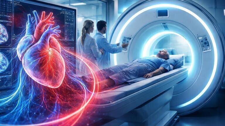
Doctors may soon be able to assess the severity of heart failure in an affected patient by using information from a routine cardiac magnetic resonance imaging (MRI) scan, according to research led by the University of East Anglia (UEA). The team has described a non-invasive method to estimate a key blood oxygen measurement that clinicians currently can only obtain via right heart catheterisation – an invasive test involving which inserts a tube directly into the heart usually via access to a vein in the leg.
Heart failure describes a clinical syndrome in which the heart cannot pump enough blood to meet the body’s needs or can do so only at the cost of raised filling pressures – whereby the pressure inside the heart is higher than normal at the point when it fills with blood between beats.
Patients can experience breathlessness, fatigue, swelling in the legs and reduced tolerance to exercise and clinicians need objective methods to measure severity, and assess the risk of deterioration and guide treatment.
In advanced disease, one important indicator is how much oxygen remains in the circulated blood that has returned to the right side of the heart. Lower oxygen values can suggest that tissues are extracting more oxygen because the blood circulation is not delivering enough which can indicate more severe haemodynamic compromise.
Right heart catheterisation can provide this information directly by measuring oxygen saturation in venous blood and by assessing haemodynamics, including pressures within the right heart and pulmonary circulation. Although the test offers valuable clinical detail, it can cause discomfort and carries its own risks, including bleeding, arrhythmia, infection and complications related to vascular access.
These concerns can weigh heavily on older patients and those who are frail or clinically unstable. This can also limit the clinicians’ ability to make repeat measurements to monitor change over time.
The investigators reported that they had developed an approach to estimate this oxygen-related measurement by using a standard cardiac MRI sequence called T2 mapping, which measures the T2 relaxation time, a property that describes how quickly MRI signal decays in tissue or blood.
In practical terms, T2 mapping creates quantitative maps that reflect how the magnetic properties of blood and tissue influence the MRI signal. Because oxygenated and deoxygenated blood differ in magnetic behaviour, the researchers said that they could use those differences to infer oxygen saturation from data gathered from an MRI scan.
“Heart failure affects hundreds of thousands of people in the UK and significantly weakens the heart’s ability to pump blood.
“Doctors often need detailed information about a patient’s circulation to decide on the best treatment,” said Professor Pankaj Garg, from UEA’s Norwich Medical School and a consultant cardiologist at the Norfolk and Norwich University Hospital, UK.
“We wanted to develop a safe, non-invasive alternative which could allow far more patients to be properly assessed and allow repeat monitoring without the risks of a catheter test,” he added.
The researchers tested the method in 30 patients, comparing the MRI-based estimates with measurements obtained from right heart catheterisation. They reported close agreement between the non-invasive estimates and the invasive readings, suggesting that the MRI approach could approximate the catheter-derived value without the need to insert a tube into the heart or to draw a blood sample for this specific measurement.
To evaluate clinical relevance, the team then analysed data from 628 people with newly diagnosed heart failure and followed them for approximately three years. They reported that participants with healthier oxygen readings on MRI had a lower likelihood of death or hospital admission related to their condition during follow-up.
Importantly, the association remained after the researchers accounted for factors that often confound risk prediction in heart failure, including age, comorbidities and overall measures of heart function. Such adjustment matters because patients with heart failure can differ widely in baseline risk and apparent predictors can reflect illness severity rather than independent prognostic value.
“One of the most important markers in advanced heart failure is how much oxygen is left in blood returning to the right side of the heart.
“Until now, getting that number has usually meant a tube test. Our study shows it can be [accurately] estimated non-invasively from a standard heart MRI,” Garg said.
The collaborators emphasised the potential practical advantages for routine care. A method that clinicians can add to a standard cardiac MRI session could, in principle, widen access because it would rely on scanning infrastructure that already exists in many hospitals, rather than on specialist catheter laboratory capacity. It could also enable clinicians to repeat assessments more easily to track disease progression and response to treatment which can be significant when the status of patients can change rapidly or when clinicians need objective evidence to support escalation of therapy.
“This [method] means we may be able to read off a crucial haemodynamic numbers from an everyday scan – effectively turning a routine MRI into a much more powerful test,” said Dr Peter Swoboda, from the University of Leeds.
Dr Gareth Matthews, from UEA, highlighted that the approach could fit within existing scanning pathways that it had potential to widen access to safer heart failure assessment across the UK’s National Health Service.
“Because this can be done as part of a standard cardiac MRI, it needs no extra hardware and no contrast dye, and adds only seconds to the scan,” Matthews added.
Despite the promise, the researchers cautioned that further work would be needed to confirm performance across different hospitals, scanner models and patient groups, and to define how clinicians should use the measurement in day-to-day decisions.
External validation matters because MRI acquisition protocols can vary between centres, and because factors such as heart rhythm, blood flow patterns and coexisting disease can influence image quality and quantitative mapping values. The team also said that future studies should assess how best to integrate the MRI-derived estimate with other established measures, such as echocardiography, blood biomarkers and clinical scoring, to support treatment selection and follow-up planning.
If validated widely, the technique could offer a safer route to quantify an important marker of advanced heart failure and could reduce reliance on invasive testing for patients who face the highest procedural risk.
For further reading please visit: 10.1016/j.jacadv.2025.102484
ILM 51.5 July 2026


.jpg)


-(1).jpg)