Laboratory products
Published over 16 years ago. See the latest and most current information on Laboratory products.
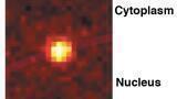
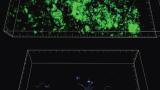
An ultrasensitive high speed camera has helped US researchers see the 3D spatial relationship between cellular structures – mitochondria and microtubules – with nanometre-scale resolution, for the first time.
Three-dimensional stochastic optical reconstruction microscopy (3D STORM) can resolve fine structural detail with a lateral (x,y) resolution of 20-30nm and axial (z) resolution of 50-60nm, using photo-activatable
fluorescent labels and a camera sensitive enough to detect single molecules.
The STORM approach uses sequential imaging of single fluorophore molecules as they toggle between bright and dark states. By exciting only a stochastic subset of single labels with an activating pulse of laser, one obtains a low light image of individual molecules that can be discerned as single diffraction-limited spots. This allows the position of each fluorescent molecule to be determined with nanometer precision. Such repeated cycles of pulses allow the position of all molecules to be determined, and subsequently the construction of a superresolution image from these precisely determined fluorophore positions.
This is the first study to use 3D STORM to visualise the spatial relationships between nano-scale structures in cells. Understanding how these structures interact paves the way for future research into cellular processes.
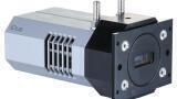
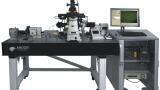
The team from Harvard University used an ultra-sensitive iXonEM+ Electron-Multiplying CCD scientific camera from Andor Technology to capture whole monkey kidney cell images from an Olympus inverted microscope. Andor’s iXonEM+ EMCCD camera is capable of detecting single photons released by the isolated fluorophore molecules.
ILM 51.5 July 2026

.jpg)


-(1).jpg)
.jpg)
.jpg)