Microscopy & microtechniques
Researchers at the Karlsruhe Institute of Technology and the University of Cambridge have developed a technique that uses a scanning laser to generate helical fluid flows, enabling the contact-free, three-dimensional rotation of microscopic particles and cells for the first time in highly viscous environments
Scientists have developed a technique that allows microscopic objects to be rotated in three dimensions without any direct physical contact, addressing a long-standing challenge in fields ranging from advanced imaging to microrobotics.
The work was led by Professor Moritz Kreysing and Dr Fan Nan of the Institute of Biological and Chemical Systems at the Karlsruhe Institute of Technology (KIT) in Baden-Württemberg, Germany, together with colleagues at the Department of Applied Mathematics and Theoretical Physics at the University of Cambridge, UK.
Precise manipulation of microscopic objects underpins many areas of science and technology, including advanced imaging, cellular biology, microfluidics, microrobotics and materials assembly. Among these capabilities, contact-free and object-agnostic three-dimensional (3D) rotation at the micro- and nano-scale has remained especially difficult to achieve.
Conventional micromanipulation methods tend to struggle in highly viscous environments, where diffusion is suppressed and force-based approaches become less effective. Many existing techniques also require specific material properties, engineered particle geometries or complex external fields which has limited their versatility.
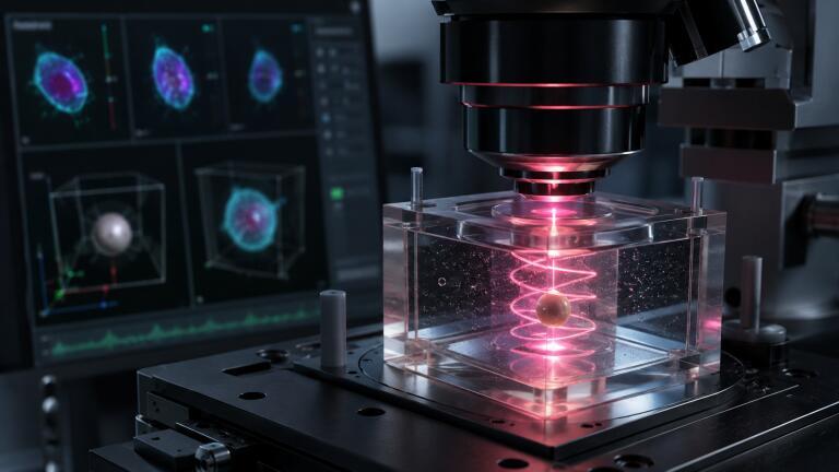
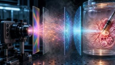
To overcome these constraints, the team introduced what it calls an opto-thermoviscous strategy which is a method that generates full 3D rotation simply by scanning a focused laser spot within a two-dimensional plane. Rather than relying on particles to act as handles or to possess particular optical properties, as in traditional optical trapping, the approach works by inducing flow directly in the surrounding viscous medium.
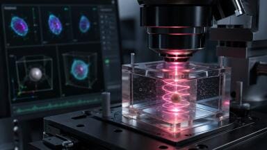
A mildly heating infrared laser is scanned rapidly across the fluid sample, and by carefully designing the scan geometry, direction and timing, the researchers were able to balance in-plane transport against out-of-plane rotational motion. The result is a fully three-dimensional helical flow capable of stable rotation, spinning, transport and trapping of a wide variety of micro-objects.
“We introduce an opto-thermoviscous strategy that scans a focused laser spot within a two-dimensional plane to robustly generate 3D helical thermoviscous flows within highly viscous media,” the authors wrote in the paper.
Through symmetry-based scan design, the team was also able to decouple rotation from unwanted lateral drift, achieving stable spinning with positional fluctuations below 200 nanometres. In the course of this work, the researchers identified a further phenomenon they term ‘opto-hydrodynamic focusing’, an effect that draws particles toward a defined height within the fluid, making the manipulation process considerably more stable and controllable.
“We further report on the discovery of opto-hydrodynamic focusing that converges a spiral motion to a defined particle height,” the authors noted.
A particular strength of the method is its material and shape independence. The team demonstrated that it works across a broad range of structures, including nano-printed tiles, stained biological cells, self-assembled particle clusters and even perfectly round, homogeneous spheres. This is notable because spherical particles are normally difficult to rotate in a controlled way, since they lack the geometric asymmetry that most rotation methods rely on.
Beyond rotation itself, the team showed that suspended assemblies could be manipulated, nano-fabricated microstructures released and reoriented, and hidden biological features exposed, including a yeast cell bud that would otherwise have remained outside the imaging plane.
One of the most promising applications lies in microscopy. Conventional 3D optical imaging suffers from markedly lower axial resolution than lateral resolution, meaning that rotating a sample into different orientations can reveal structural detail that would otherwise remain obscured.
By combining stepwise particle rotation with volumetric microscopy and multi-view image fusion, the researchers demonstrated the technique’s imaging potential in proof-of-concept experiments on HCT116 cells, a widely studied human colon cancer cell line. This approach successfully resolved two distinct cell nuclei that could not be separated in a conventional single-view confocal dataset, thereby improving the effective 3D imaging resolution achievable with existing microscopes.
The authors described the broader significance of the findings in the paper: “Conceptually, this elevates thermoviscous flows from planar transport to symmetry-engineered volumetric actuation, delivering robust, material-agnostic, out-of-plane rotational control and sheathless hydrodynamic focusing for all-optical micromanipulations and augmented microscopy.”
They added that the work establishes: “a novel paradigm in microscale flow control for micromanipulation in the life and engineering sciences and constitutes a significant step toward a future of generally applicable 3D microrobotics.”
By turning high viscosity, usually a hindrance to micromanipulation, into an advantage, the technique offers a powerful new route to precise 3D control of arbitrary micro-objects in environments where conventional approaches perform poorly. The researchers suggest the method could find future application not only in biological imaging, but also in next-generation microrobotic systems and advanced microscopy workflows more broadly.
For further reading please visit: 10.1038/s41377-026-02303-8
ILM 51.6 Sept 2026


 Product directory


.jpg)
.jpg)
















.jpg)