Raman
A novel method for fabricating uniformly structured gold nanostructures has raised the accuracy of label-free cell identification from 75 per cent to 96.2 per cent, offering a more reliable route to sorting cancer and non-cancer cells without biomarker labelling
Cell sorting is a foundational step in precision biomedicine, separating mixed populations of cells into pure subgroups based on their distinguishing features. Blood and tissue samples typically contain many cell types and this mixture can obscure the signals that genetic or protein-based tests are trying to detect.
Sorting cells into uniform groups allows researchers to study cell function, stem cell development and disease mechanisms with greater clarity, and supports drug screening and quality control of cell-based products. The analysis of rare cells, such as circulating tumour cells and cancer stem cells, is attracting growing interest because of its relevance to drug development and clinical diagnostics.
Flow-cytometry-based sorting was first described in the 1960s, and fluorescence-activated cell sorting and magnetic-activated cell sorting remain the most widely used methods today. Both pass single cells through laser detection systems, before charged droplets containing target cells are deflected by an electric field for collection.
To avoid the need for labelling and invasive manipulation, researchers have more recently developed label-free alternatives, including image-activated cell sorting, acoustics-activated cell sorting and Raman-activated cell sorting. These systems identify and sort cells using intrinsic properties such as shape, acoustic characteristics and the molecular composition captured in a Raman spectrum. However, their sorting accuracy is limited in complex cell populations.
Deep-learning approaches based on neural networks have since been applied to improve identification accuracy but such algorithms are computationally demanding and often impractical for untrained users. Low sensitivity and limited spatial resolution remain the main barriers to accurate label-free cell sorting, since both directly affect the accuracy of the entire process, from identification through to isolation.
Surface-enhanced Raman scattering (SERS) offers an effective, non-invasive route to identifying single-molecule fingerprints in biosensing, with ultra-sensitive detection and analysis. Because Raman signals are highly accurate and quick to acquire, SERS can support cell sorting more efficiently than image- or acoustics-based methods, and Raman mapping's spatially resolved signals help reduce overall processing time. Deep-learning-based SERS techniques for cell identification have followed, with earlier studies using gold nanoparticle substrates to collect cancer-cell signals for neural-network models used in early cancer diagnosis. Even so, selecting the right Raman tags or biomarkers remains critical to accurate SERS analysis and deep learning alone is not a universal solution for cell identification.
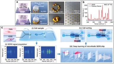
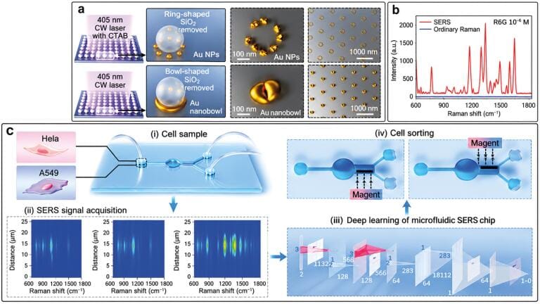
Now, the research group of Professor Koji Sugioka and Dr Shi Bai, from the Advanced Laser Processing Research Team at RIKEN, part of the RIKEN Center for Advanced Photonics in Wako, Saitama, Japan, has developed a new method for fabricating a SERS substrate with uniformly distributed hotspots, using laser near-field reduction of gold ions.
“This technique allows the creation of plasmonic ring-shaped nanostructure arrays by introducing cetyltrimethylammonium bromide into the precursor solution,” said Sugioka.
The gold nanoparticles produced are around 28 nanometres in size, while the number of nanoparticles in each ring structure – and the gaps between them – depend on the reduction time which is a factor the team found critical to consistent SERS performance.
Raman mapping showed that the ring-shaped nanostructure arrays produced highly uniform Raman enhancement across the surface, with a relative standard deviation of two per cent. The arrays also achieved high spatial resolution, around 185 nanometres, confirmed experimentally through Raman mapping. This spatial resolution proved essential to deep-learning-based, label-free cell identification with the higher resolution of the ring-shaped arrays raising identification accuracy from 75 per cent to 96.2 per cent, which suggested spatial resolution should be a key consideration in future work of this kind.
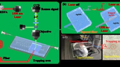
As a proof of concept, the researchers used femtosecond laser processing to fabricate a cell-sorting device, successfully separating cancer and non-cancer cells within a ‘Y’-shaped microchannel.
“The preliminary results indicate that the developed sorter achieves precise cell sorting and separation without the need for biomarker or Raman-tag labelling,” concluded Sugioka.
For further reading please visit: 10.29026/oea.2026.260071
ILM 51.6 Sept 2026



 Product directory