Microscopy & microtechniques
A study has linked reduced fungal viability with changes in cell-wall chemistry, surface structure and nanoscale mechanical behaviour through a combination of complementary laboratory techniques
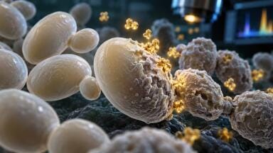
A research team drawn from universities in Poland has used a combination of cell analysis, microscopy and spectroscopy to examine how a quercetin-derived complex affects Candida albicans. The work has connected a concentration-dependent loss of viability with changes in cell-wall organisation, surface shape and mechanical properties, which provides a broader picture than an antimicrobial activity measurement alone. Candida albicans is both a normal member of the human microbiome but, importantly, an opportunistic pathogen.
The study investigated the sodium complex of quercetin-5′-sulphonic acid. The experiments assessed the compound’s effects in vitro, with an emphasis on the physical and chemical alterations that accompanied exposure.
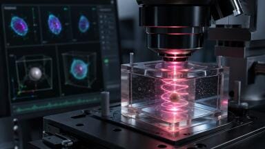
Flow cytometry indicated that viability declined as the concentration of the complex increased. In this technique, individual cells pass through an optical measurement system so that signals from many cells can be assessed. The resulting distribution can reveal differences within a population that a single average measurement might conceal, although interpretation depends on the assay’s markers and controls.
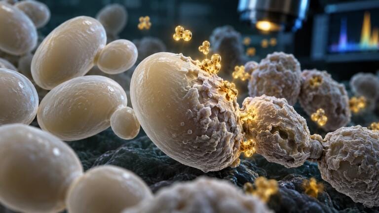
The researchers complemented this assessment with Calcofluor White and Congo Red stains to examine cell-wall organisation. The treated cells showed structural alterations and incomplete separation of daughter cells. That latter observation is relevant because division and physical separation require coordinated changes to the cell wall. A disturbance can therefore affect both the appearance of individual cells and the organisation of the population.
However, these observations do not identify a specific molecular target on their own. A change in cell-wall appearance may arise from direct interference with wall components, disruption of the processes that maintain them, or broader cellular damage. The value of the additional analytical methods was to establish whether other forms of evidence supported the structural changes and helped describe their nature.
Scanning electron microscopy confirmed deformation at the fungal surface. This method supplies detailed images of external morphology and can reveal features that are difficult to resolve through routine light microscopy. Its findings supported the view that exposure had affected the physical cell envelope, although specimen preparation and image selection remain relevant to interpretation.
Atomic force microscopy then added measurements at a smaller scale. A fine probe interacts with the sample surface to assess topography and force response. The researchers recorded increased surface roughness, elasticity and deformability after treatment. These parameters described how the cell surface appeared and responded mechanically under the particular measurement conditions.
Mechanical properties require careful distinction with roughness concerning variations in surface height, while deformability concerning the extent to which a sample changes shape under force. Elastic behaviour pertains to its response regarding deformation and recovery. These measurements can help characterise an altered cell envelope but they should not be collapsed into an unqualified claim that a cell has become stronger, weaker or more resistant to rupture.
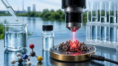
Fourier-transform infrared spectroscopy provided a chemical counterpart to the microscopy. The technique measures infrared absorption associated with molecular vibrations. Changes in a spectrum can indicate differences in the chemical composition or environment of a sample. In this study, dose-dependent spectral changes were consistent with altered cell-wall chemistry after exposure to the quercetin complex.
The combination of methods is the principal analytical strength with flow cytometry addressing viability, staining highlighted wall organisation, electron microscopy examining morphology, and atomic force microscopy as an assessment of nanoscale topography and mechanics, with infrared spectroscopy supplying chemical information. Agreement between these different forms of observation made the overall account more informative than any single result would have been.
However, complementary observations do not automatically establish a sequence of cause and effect. A damaged cell wall could contribute to reduced viability, while a cell that has lost normal metabolic function could also develop secondary structural changes. Experiments that resolve the timing of each event, together with targeted biochemical measurements, would be needed to distinguish those possibilities more firmly.
The results also concern one organism and one compound under controlled laboratory conditions. They do not establish that the same effects would occur in other fungal species, in a mixed microbial community or in an infection. The chemical environment around a cell can alter a compound’s availability, while the architecture of an organised fungal community can change exposure relative to that of freely suspended cells.
Selectivity remains a central question for any antimicrobial candidate. Activity against fungal cells must be considered alongside effects on human cells at relevant concentrations. The present experiments did not establish an acceptable therapeutic window, suitable pharmacological exposure or efficacy in an infection model. The quercetin origin of the complex does not answer those questions, since chemical modification and formulation can alter biological behaviour.
For microscopy and spectroscopy laboratories, the study offers an example of how to connect antimicrobial observations with several independent dimensions of cell damage. Its findings support further investigation of the complex and of the cell-envelope processes affected by exposure. The wider methodological lesson is that viability, chemistry and mechanics provide distinct evidence, and that a convincing account of antimicrobial action requires their relationships to be tested rather than assumed.
For further reading please visit: 10.1038/s41598-026-69531-1
ILM 51.6 Sept 2026


 Product directory


.jpg)
.jpg)















.jpg)