Laboratory products
Published over 16 years ago. See the latest and most current information on Laboratory products.
Introduction
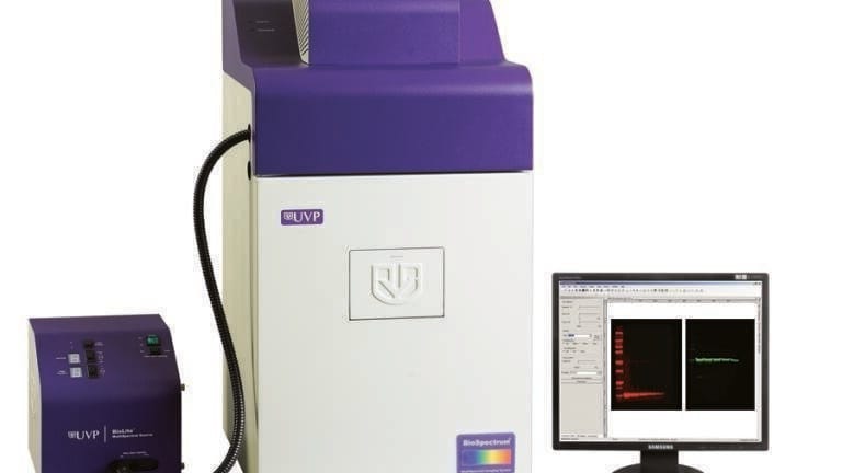
The BioSpectrum combined with the BioLite MultiSpectral Light Source provide a powerful system to excite and illuminate wavelengths for multiplex NIR imaging. The BioSpectrum System provides precision NIR filters for excitation, and also offers rapid, high resolution image capture through the use of cooled CCD camera and low light lenses. Exposures times are very fast, typically less then 2 minutes; much faster than laser scanning.
Researchers can detect and quantify virtually any NIR dye. Protein blotting determines the presence or absence of one or more proteins and can provide additional information on quantity of protein (Gallagher and Wiley, 2008).
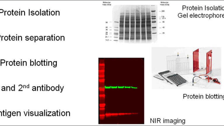
Use SDS PAGE (Gallagher and Wiley, 2008) to first separate the proteins by size, followed by electrotransfer of proteins onto nitrocellulose or PVDF membranes (Figure 1).
Figure 1. The immunoblotting process. Protein blotting starts with SDS PAGE separation followed by blotting and antibody probing and analysis.
The membrane surface absorbs the protein, and the researcher probes the membrane with primary antibodies specific to the protein of interest. Enzyme or fluorescent dye tagged secondary antibodies are used to identify the primary antibody binding site. If only one protein species is being identified, then only one primary antibody and label is used. If multiple proteins are being identified on the same blot, then the analysis relies on probing each protein with a different primary antibody and secondary antibody with a different fluorescent tag, yielding a multiplexed result.
Detection of the primary antibody binding to the protein of interest is accomplished via a fluorescent label attached directly to the secondary antibody.
Imaging
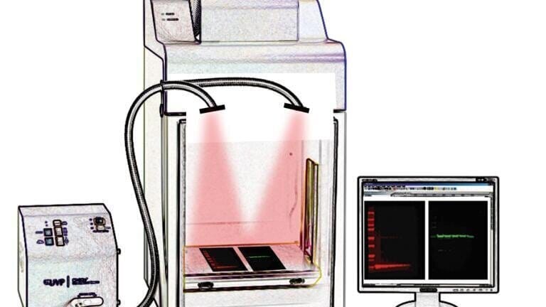
For fluorescent imaging, the membrane is illuminated with overhead monochromatic excitation light from the BioLite MultiSpectral Source. The induced fluorescence at each of the tagged sites is captured with a cooled CCD camera through an emission filter in the BioSpectrum that selects for the fluorescent light while blocking excitation light (Figure 2). Images were processed with VisionWorks® LS image acquisition and analysis software (UVP, LLC).
Figure 2. Together the BioSpectrum and BioLite excite and illuminate NIR. Up to 8 excitation and 5 emission wavelengths are possible in a single experiment.
Discussion
Figure 2 illustrates the NIR multiplex illumination capabilities of the BioSpectrum and BioLite system, which clearly and specifically separate NIR emission wavelengths of 680 and 800 nm. NIR labels for protein blot applications offer many advantages. They permit multiplexing so that several proteins in a sample can be detected and analyzed simultaneously on a single protein blot. NIR labels offer very low background and high signal-to-noise ratio for quantitative imaging and include high stability of the label on the processed and dried blot.
Conclusion
Routine NIR imaging with the BioSpectrum and BioLite MultiSpectral Source is fast, efficient, and straightforward, yielding full high definition images for quantification and publication.
References
Gallagher, S.R. and Wiley, E.A. Current Protocols: Essential Laboratory Techniques. Wiley, 2008
Link: http://j.mp/YghY1o
ILM 51.5 July 2026


-(1).jpg)
.jpg)


.jpg)
.jpg)