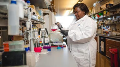
Research news
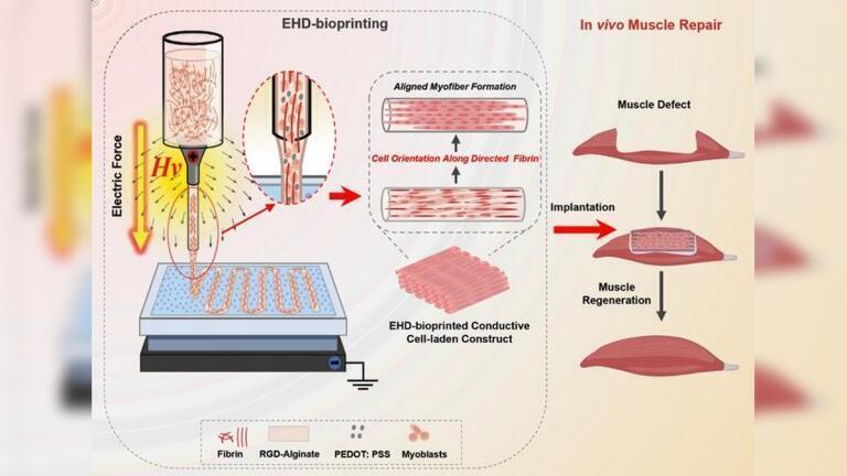
Researchers at Xi’an Jiaotong University have reported a way to bioprint three-dimensional skeletal muscle that combines realistic shape with the internal cellular alignment that gives muscle its strength. The approach uses an electric field during electrohydrodynamic bioprinting to reorganise fibrin within a bioink into nanoscale guidance fibres, which then invokes muscle cells to align, fuse and mature
Building functional human skeletal muscle in the laboratory has remained a central ambition in regenerative medicine, partly because muscle function depends on more than tissue bulk or outward form. In native muscle, myogenic cells fuse into long fibres that are organised in a highly ordered manner, and the direction of the fibres shifts from one muscle to another to match biomechanical demands. Tissue engineers have learned to shape living constructs in three dimensions but it has been difficult to reproduce the internal order such that muscle can contract with both force and efficiency.
A team at Xi’an Jiaotong University, Xi'an, China, has now reported a strategy to address both external geometry and internal organisation in a single fabrication step. The researchers have described how they used electric forces during electrohydrodynamic bioprinting to create muscle-like constructs in which embedded cells aligned along well-defined directions, including straight, curved and circular patterns.
The study has suggested that an electric field can do more than guide deposition with high precision. It can also help to ‘instruct’ cells indirectly, by reorganising material features within the printed filament that cells then interpret as structural cues.
Skeletal muscle architecture varies markedly across the body. Many muscles that drive gross movement, such as those in the limbs, contain fibres arranged in broadly parallel bundles. Others show more complex geometries, including curved or fan-like arrangements that support fine control in functions such as mastication or gripping.
Despite this diversity, muscle shares a common microscopic principle, in that myogenic cells must align in order to fuse into elongated myofibres, assemble contractile machinery and transmit force along a coherent axis. When alignment fails, tissue remain mechanically weak and physiologically immature.
Conventional tissue-engineering approaches have supported alignment in simplified settings, often in two-dimensional sheets or thin scaffolds in which surface features, mechanical strain or micropatterns can bias cell orientation. Three-dimensional bioprinting can fabricate bulk shapes and spatially varied structures but cells within printed hydrogels often remain comparatively disordered.
“You can print a muscle-like shape but its cells don’t know in which direction to pull,” said Professor Jiankang He, the study’s corresponding author and a professor of mechanical engineering at Xi’an Jiaotong University.
To overcome this mismatch, the researchers used electrohydrodynamic bioprinting which differs from more familiar extrusion-based bioprinting. Rather than push a viscous bioink through a nozzle with pneumatic or mechanical force, electrohydrodynamic bioprinting applies a strong electric field that draws out an ultrafine jet from a liquid meniscus.
This mechanism can deliver markedly higher resolution but previous implementations have offered limited leverage over how cells organise themselves after deposition, particularly within thicker three-dimensional constructs.
The key change in the present work lay in the composition and behaviour of the bioink under an electric field. The team combined alginate, a widely used printable hydrogel, with fibrin, a fibrous protein central to blood clotting and wound repair. Fibrin also responds to electrical and mechanical cues and the researchers exploited that property during printing.
Under high voltage, the liquid at the nozzle tip forms a characteristic meniscus known as a ‘Taylor cone’, from which the jet emerges. The authors reported that, at approximately 3,000 volts, small and initially disordered fibrin aggregates within the bioink elongated and reorganised into evenly aligned nanofibres oriented along the axis of the printed filament.
Those nanofibres then acted as internal guidance structures so that the cells embedded in the hydrogel and then sensed the anisotropic environment, so orienting their cytoskeleton accordingly, which helped to align cell bodies and encourage fusion into longer muscle fibres.
“As the material aligns, the cells follow,” said Ayiguli Kasimu, the study’s first author and a doctoral candidate.
“The electric field is effectively building a road system at the nanoscale, and the cells naturally grow along it,” she said.
Because this alignment emerged during deposition rather than as a post-printing conditioning step, the method allowed the researchers to programme different fibre directions by altering the print path. By changing the trajectory of the nozzle, they produced constructs with linear alignment, curvature or circular organisation while maintaining tight internal order. That capacity matters because muscle function depends on architecture. A construct that matches the gross shape of a target muscle but fails to replicate its directional anisotropy will not reproduce its native mechanics, even if the cell types are appropriate.
The researchers also aimed to support the electrical aspects of muscle physiology. Skeletal muscle relies on electrical excitation to coordinate contraction and poor conductivity in hydrogels can limit maturation in engineered tissue. The team therefore incorporated conductive polymers into the bioink to improve signal transmission within the constructs.
“Muscle tissue relies on electrical signals to coordinate contraction, and the conductive additives allowed the printed constructs to transmit these signals,” said Assistant Professor Zijie Meng, a co-corresponding author at Xi’an Jiaotong University.
The authors reported that the conductive, aligned environment supported more efficient fusion into mature fibres and enhanced expression of muscle-associated proteins which are commonly used as markers of myogenic differentiation and functional maturation.
To assess performance in vivo, the researchers implanted the printed constructs into animal models with muscle defects and reported improved functional recovery alongside evidence of new muscle formation. In that setting, the constructs appeared to do more than persist after implantation. They contributed to tissue restoration in a way consistent with the intended design principle, namely that aligned, electrically competent scaffolding can guide organised regeneration, rather than merely fill space.
Beyond muscle repair, the work has advanced a broader concept in biofabrication through the use of electric fields as internal patterning tools. In the authors’ account, alignment arose from coupled electrical and mechanical effects. Electrical forces promoted migration and reorganisation of fibrin components, while intense stretching during jet formation elongated fibrin clusters into directional fibres. Together, these processes produced a microenvironment that cells could interpret without the need for added micropatterned moulds or prolonged external conditioning.
The molecular details that govern fibrin’s response to an electric field remain incompletely resolved and further optimisation will be needed to tune variables such as cell density, material formulation and longer-term stability and function after implantation. Even so, the study has offered a clear demonstration that electrohydrodynamic bioprinting can couple high-resolution fabrication with emergent biological order.
By treating the electric field as a design signal rather than merely a printing aid, the Xi’an Jiaotong University team has outlined a route to bioprint living skeletal muscle that better reflects native structure and function.
For further reading please visit: 10.1088/2631-7990/ae3923
ILM Guide 2026/27


.jpg)


2.jpg)
